Hip Fractures Fixation with Nails – Everything you need to know

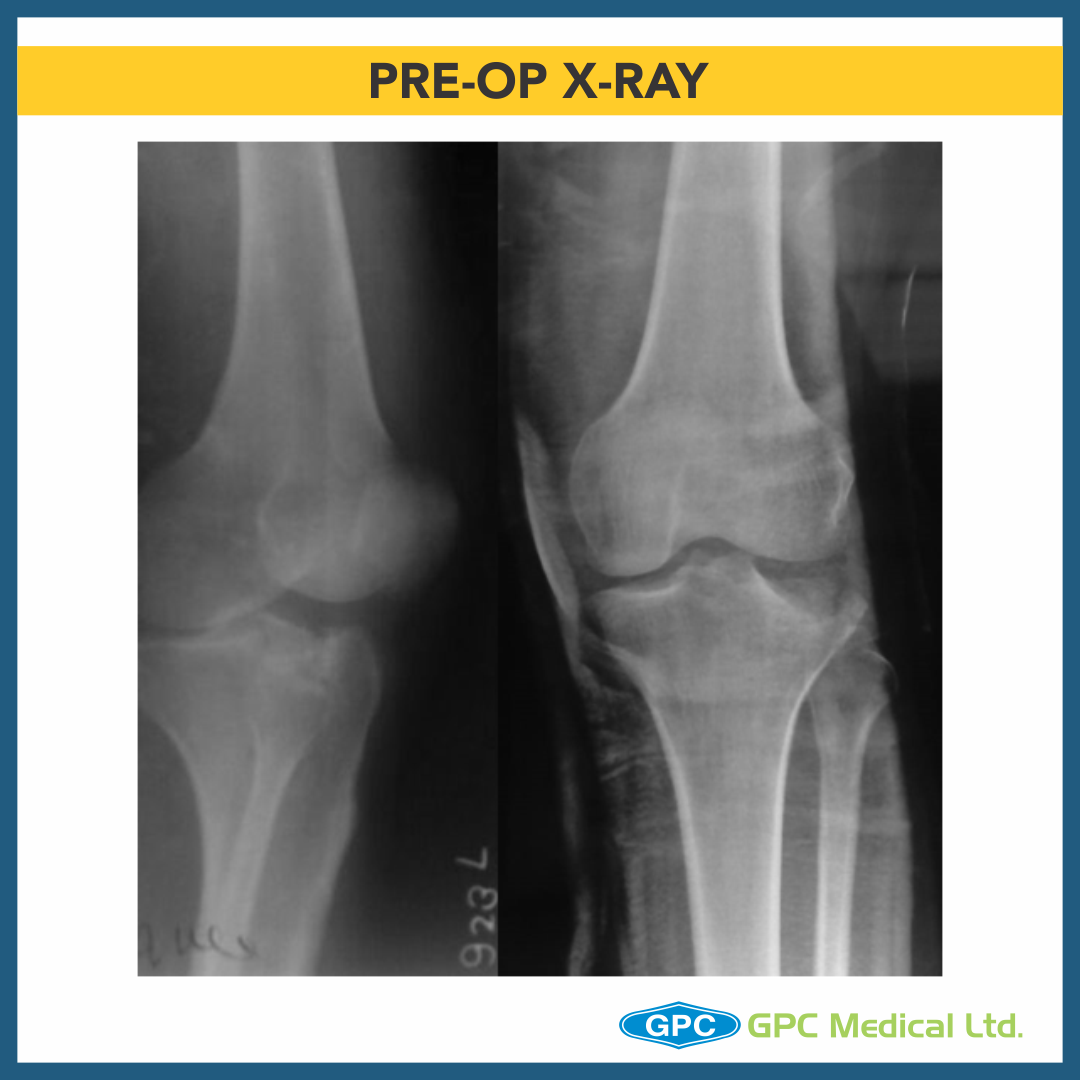
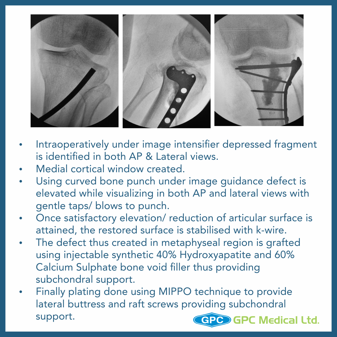
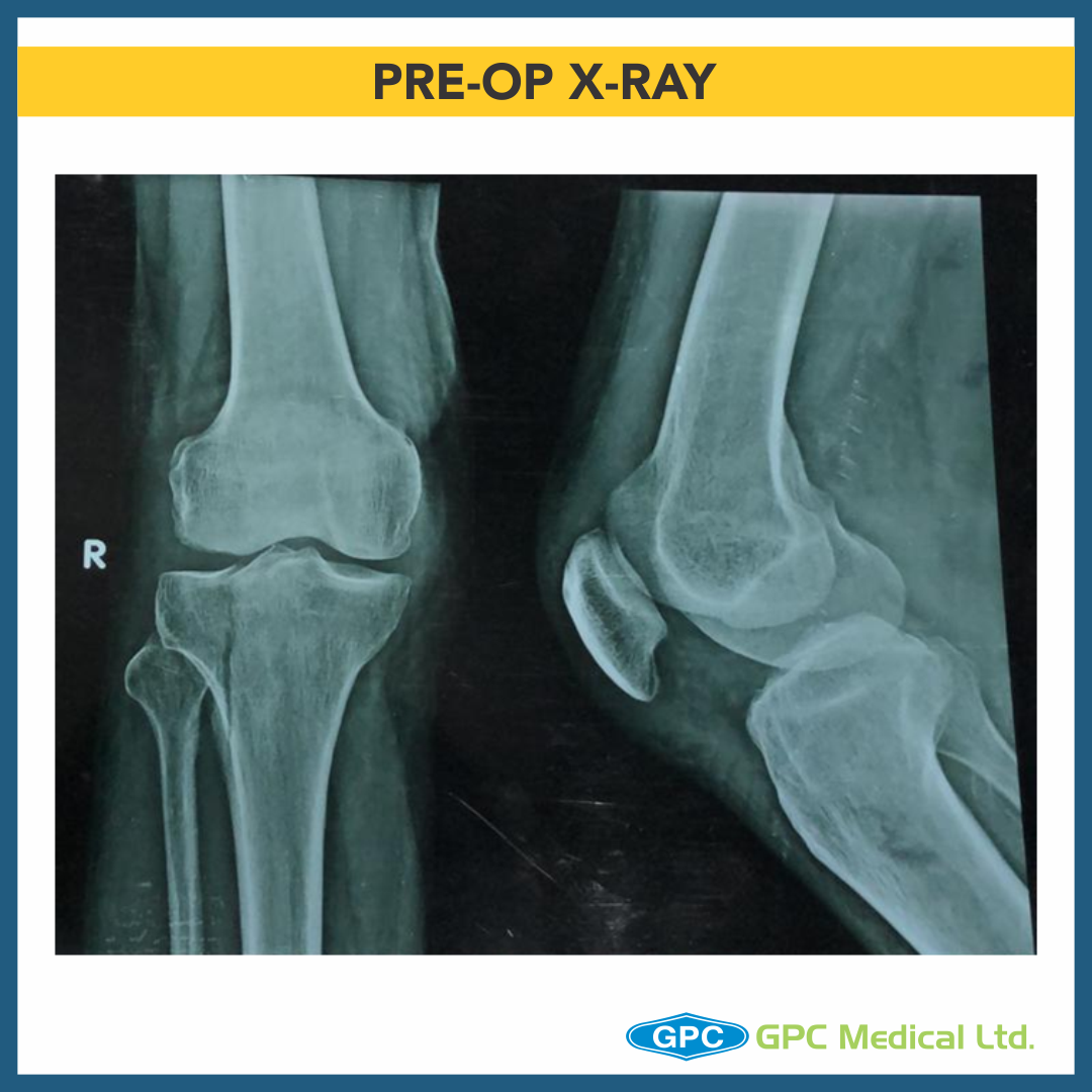
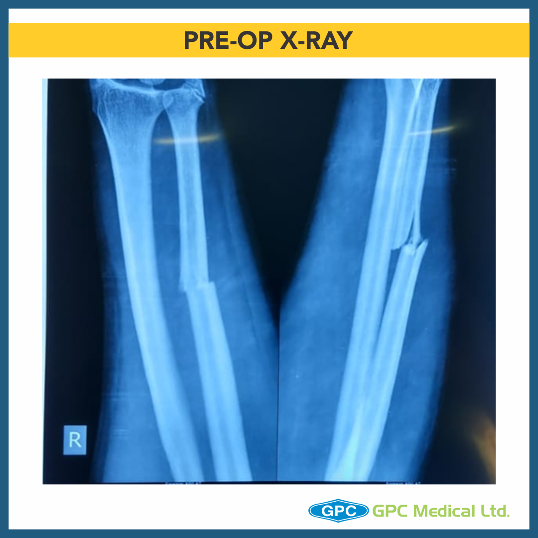
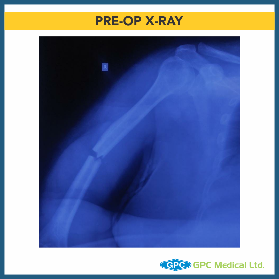
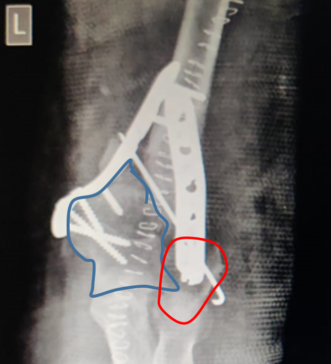
According to a survey, the number of hip fractures is expected to surpass 6 million worldwide by the year 2050. Hip fractures are breaks in the upper portion of the femur that usually occurs in elderly patients whose bones have become weakened by osteoporosis. In case of younger patients, hip fractures occur due to a high-energy event, such as a fall from a ladder or vehicle collision. Most of these fractures occur in older patients who are injured in household or community falls.

Hip fractures tend to be very painful. This is why prompt surgical treatment is recommended. Treating the fracture and getting the patient out of bed as soon as possible helps prevent medical complications later on such as bed sores, blood clots and pneumonia. Disorientation can also occur in very old patients due to prolonged bed rest which makes rehabilitation and recovery much more difficult.

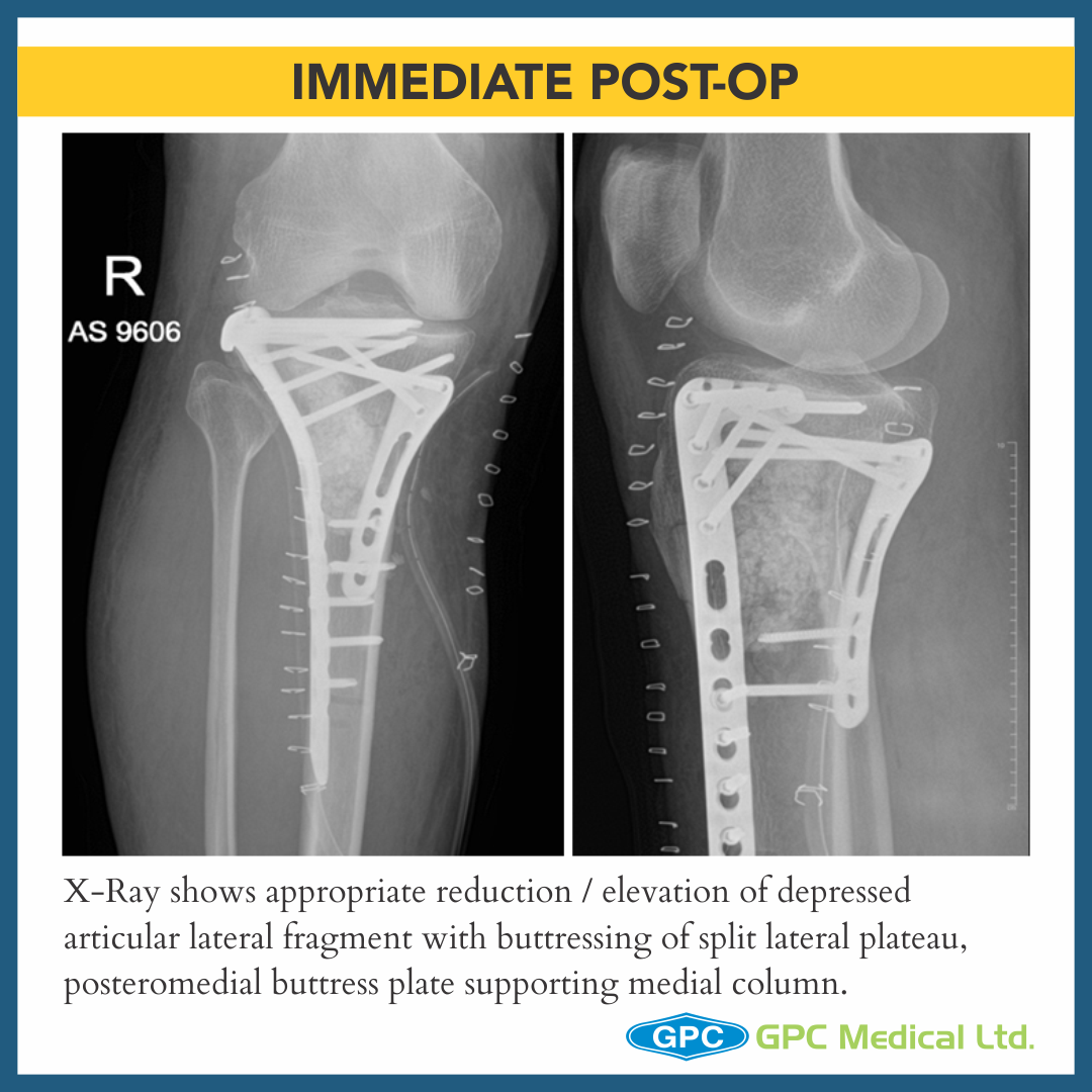
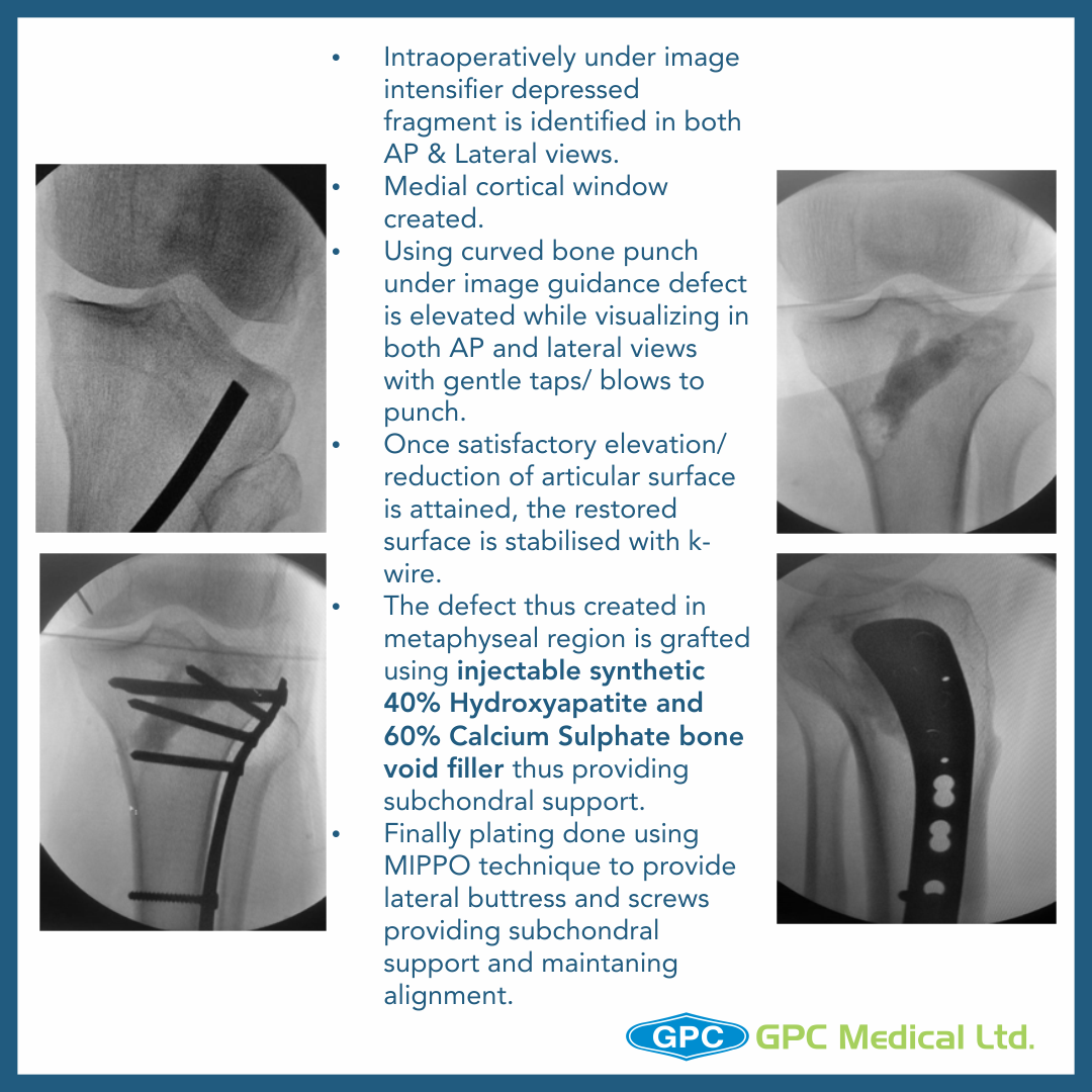
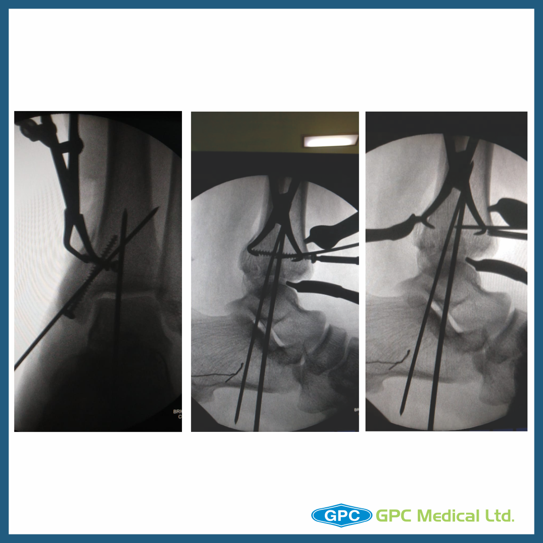
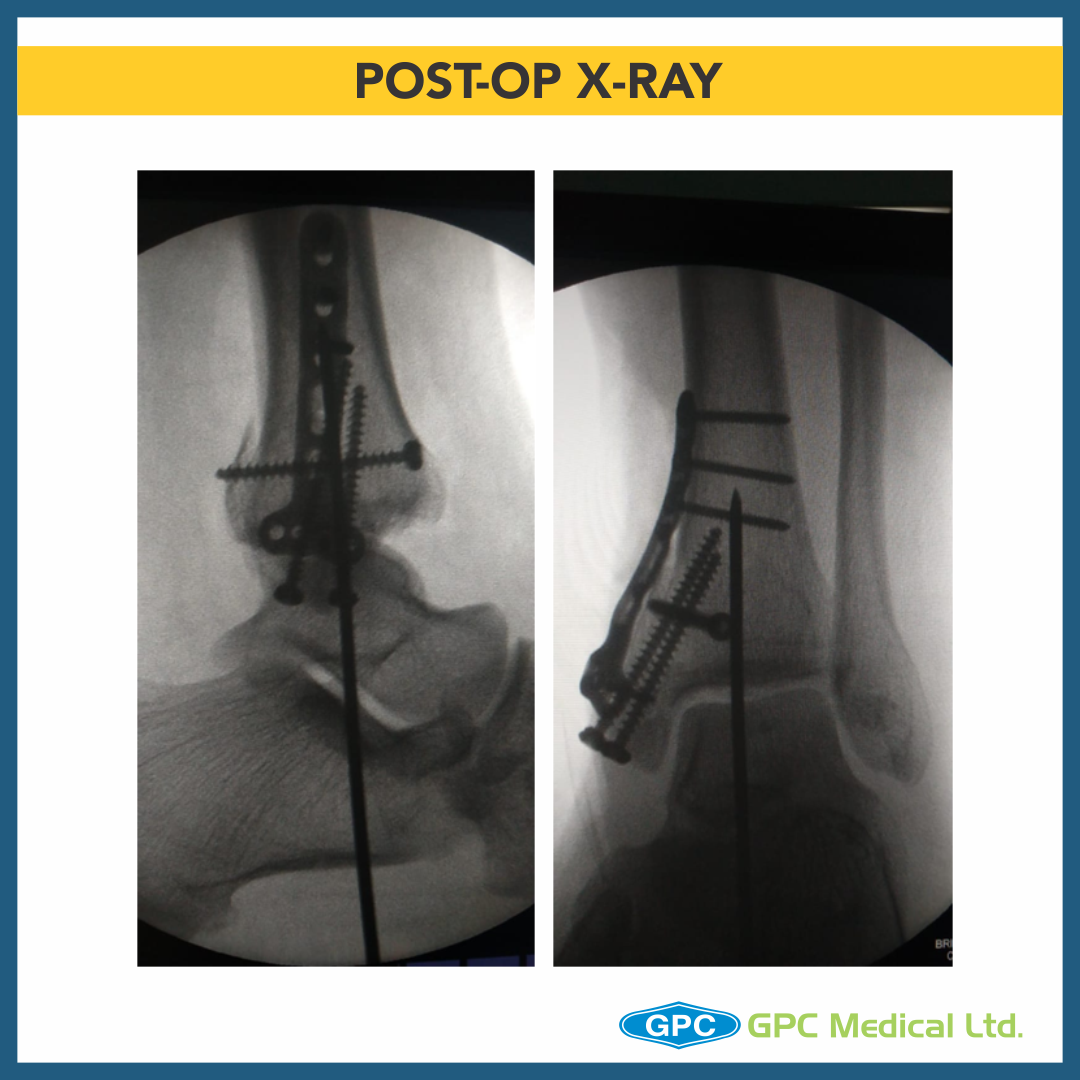
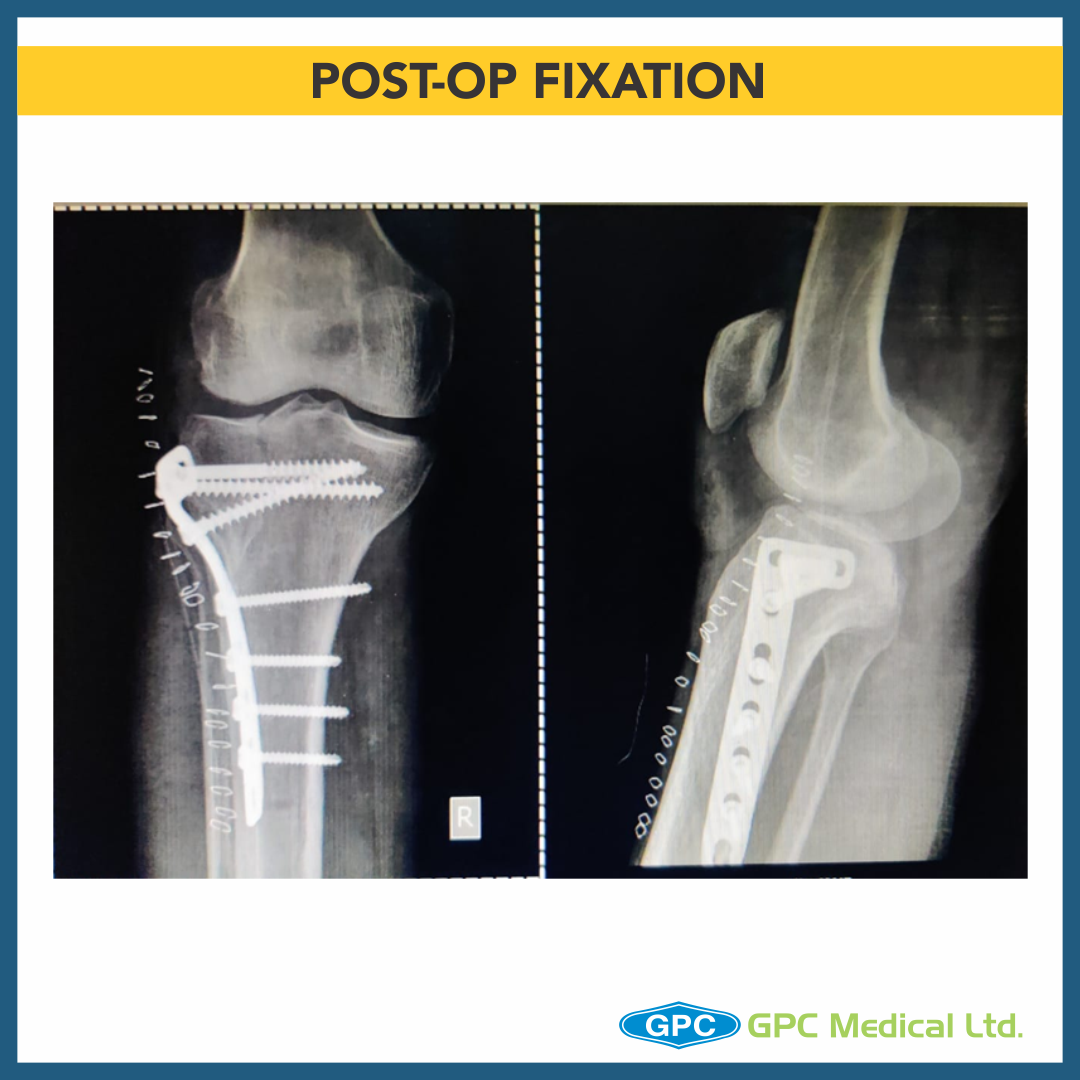
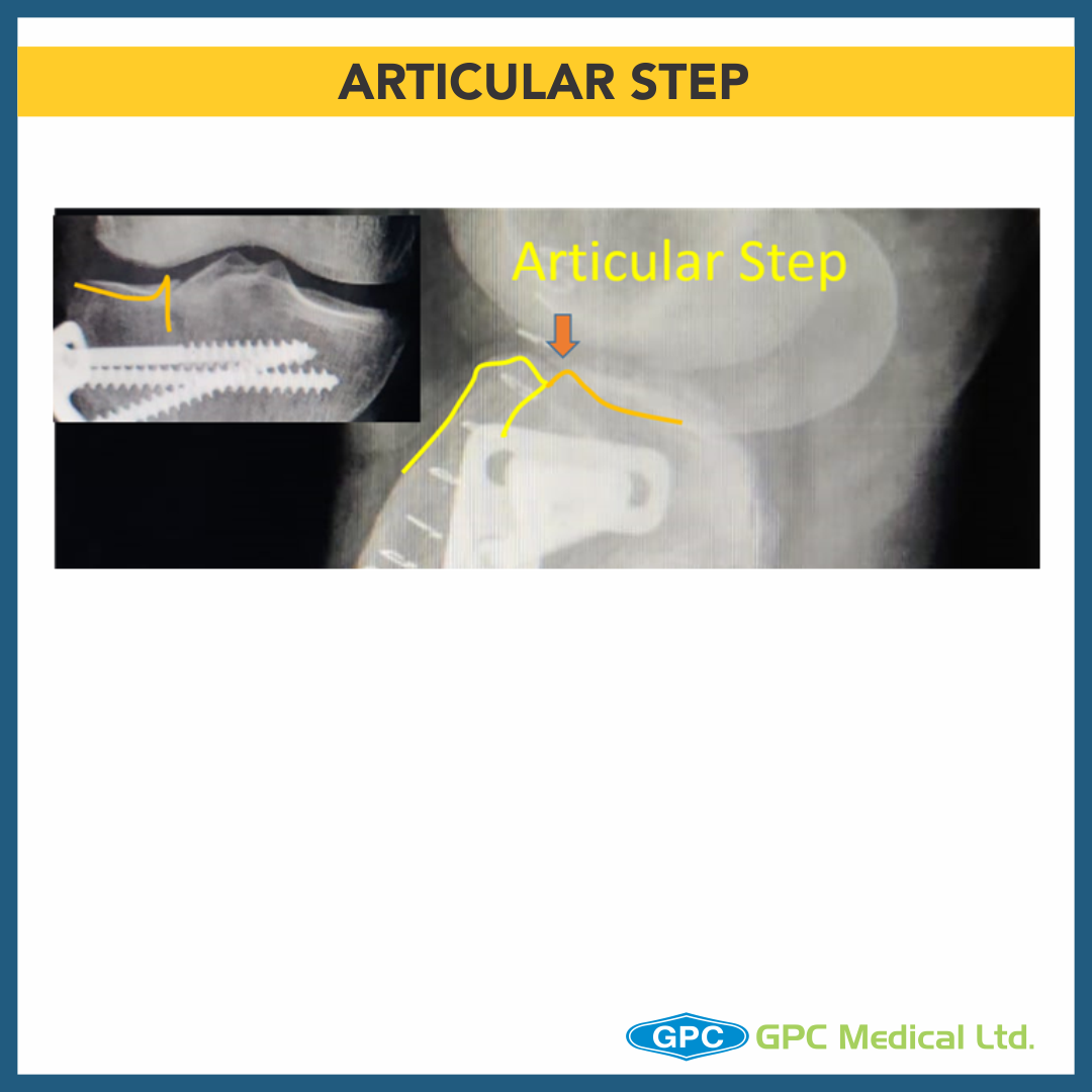
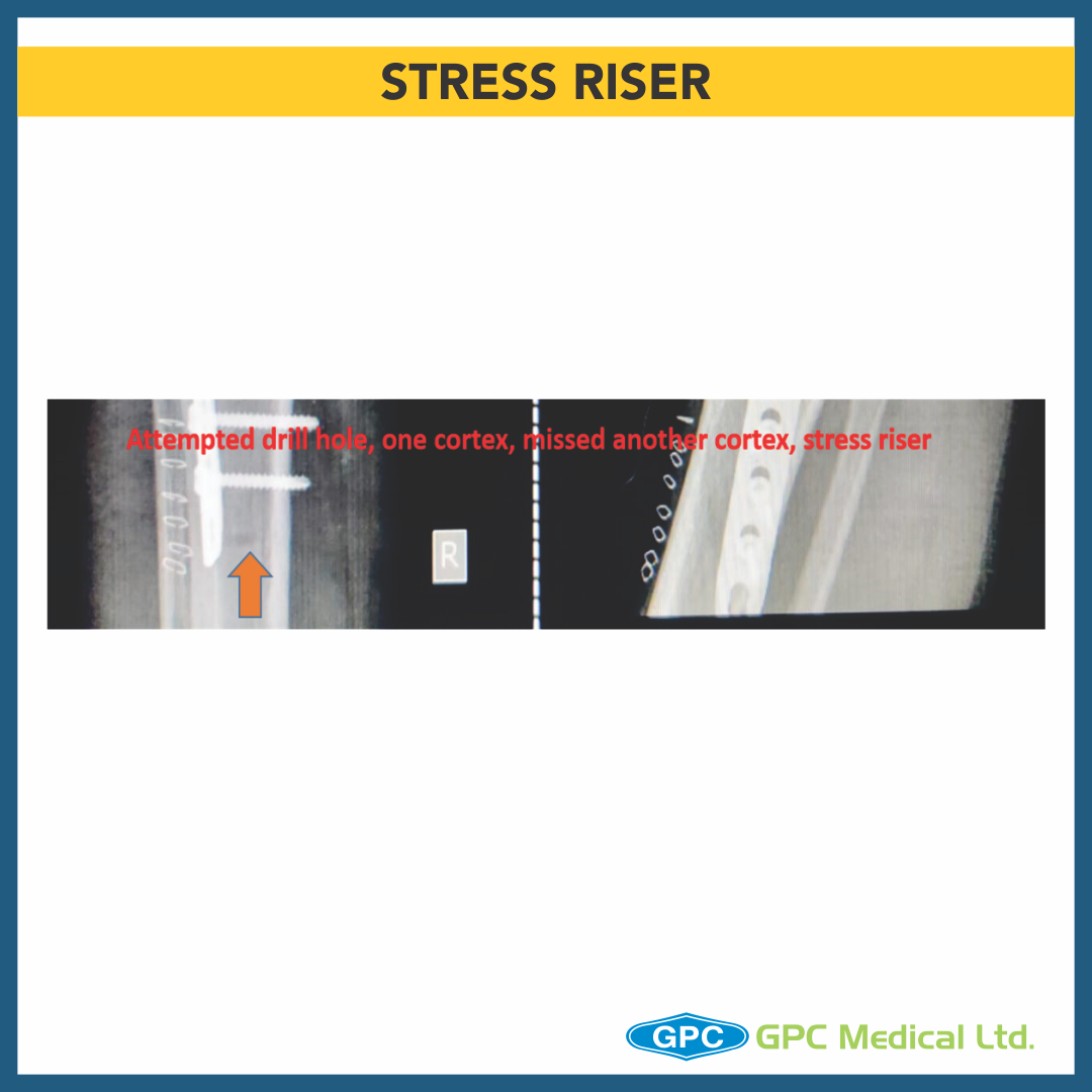
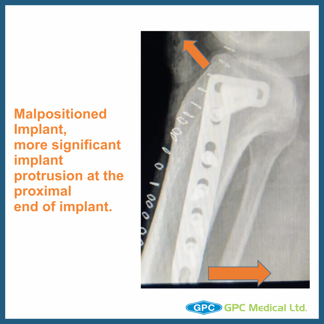
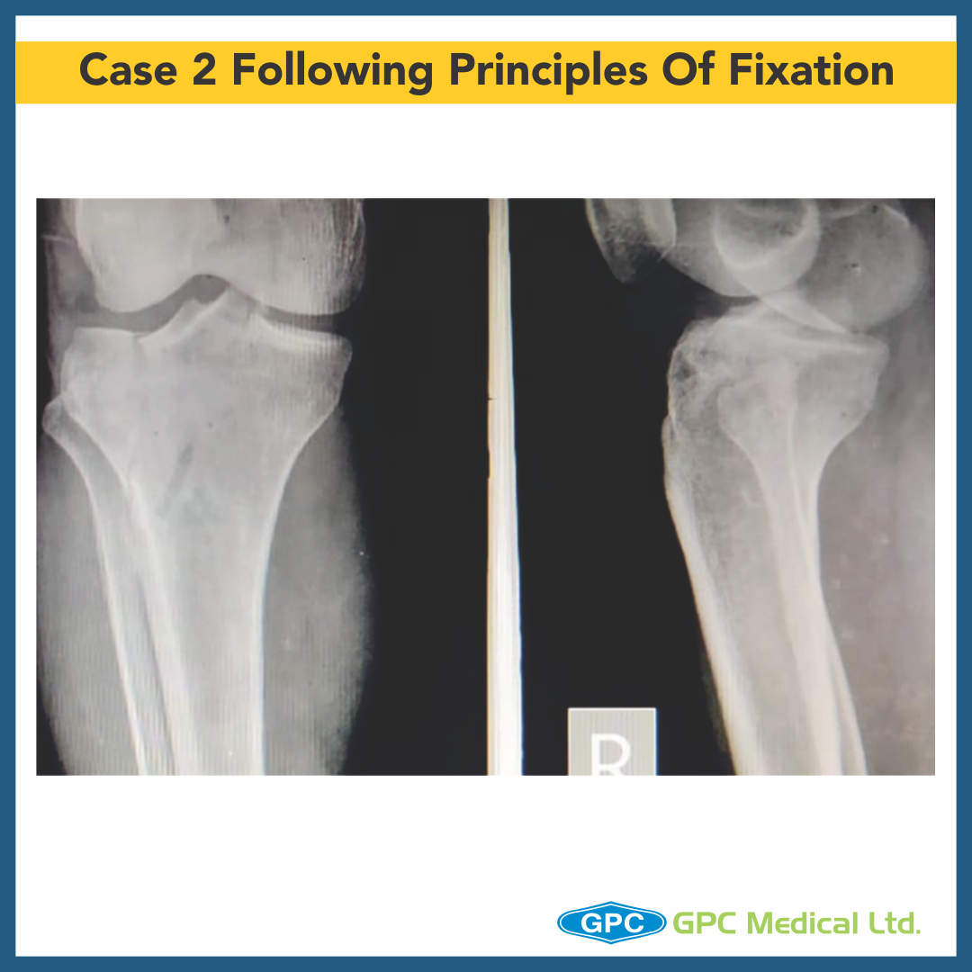
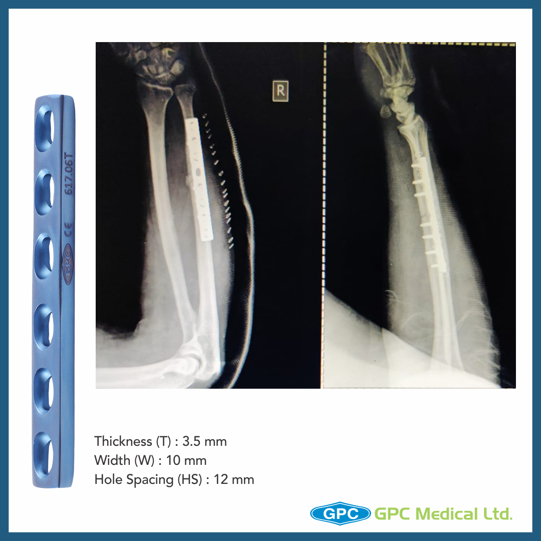
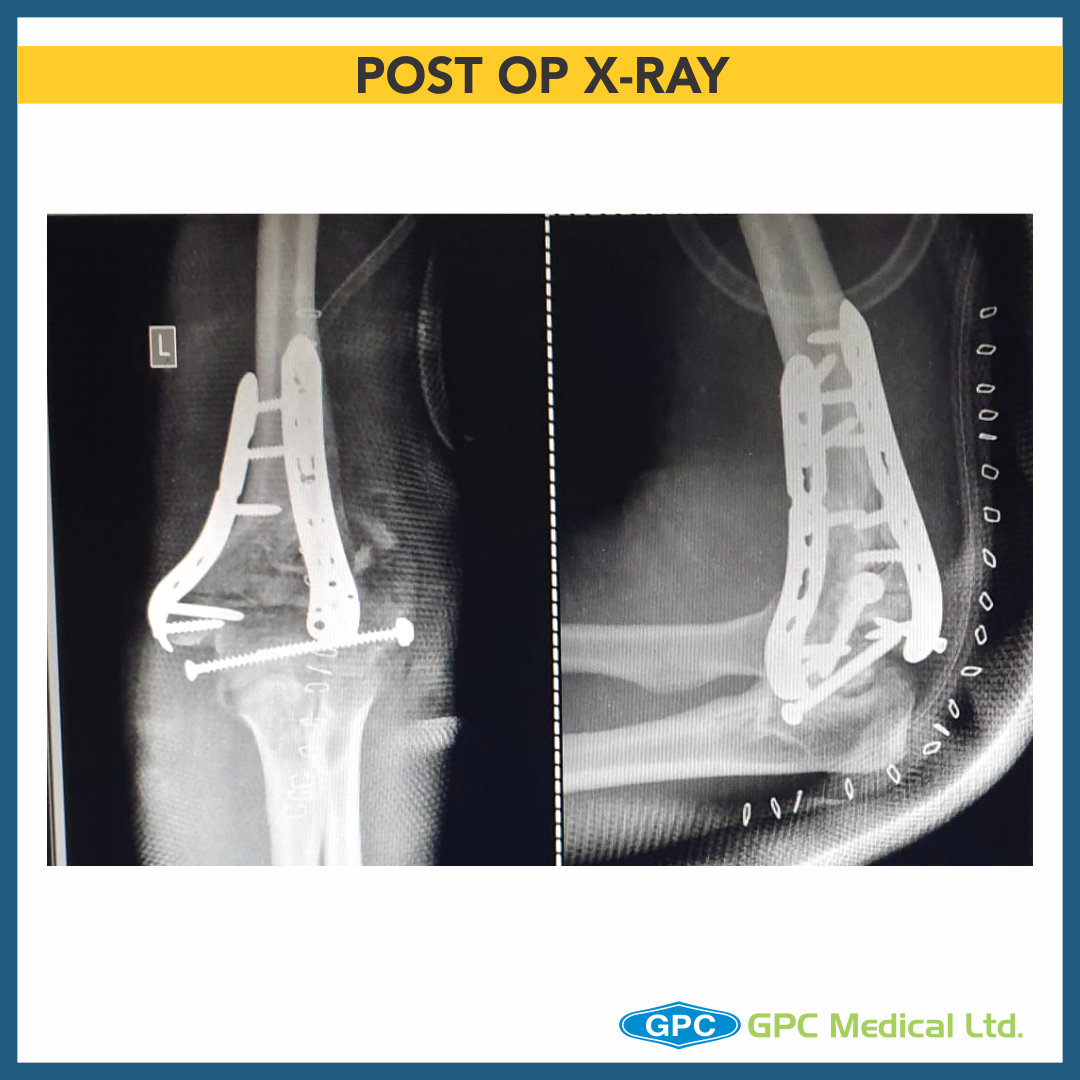
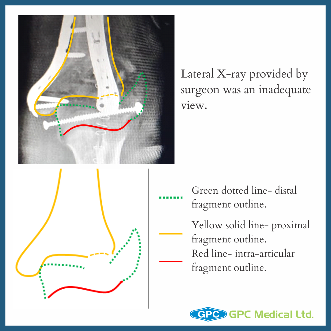
Orthopedic implants like nails play an important role in the fixation process. The choice of the implant material is very crucial as it influences rigidity, corrosion, bio-compatibility and tissue receptivity. The surface morphology of the implants also affects its stability within the skeleton or the surrounding cement mantle.

GPC Medical has been serving the medical world as a leading manufacturer of medical equipment and healthcare systems for more than 20 years. As one of the top manufacturers and suppliers of medical equipment and affordable healthcare solutions in India, GPC Medical strives continuously to innovate medical solutions that enriches the lives of people. We are an ISO 9001, ISO 13485 certified company that exports and supplies medical equipment and surgical instruments worldwide.

GPC Medical manufactures a wide range of nails for hip fractures that are made from the best raw materials with ergonomic design. Let’s take a look at the various types of hip nails:

intraHEAL Proximal Hip Stabilizing Nail (ILBS59)

Our IntraHEAL Proximal Hip Stabilizing Nail is the best-in-class nail in the market for intertrochanteric fractures of femur. Our IntraHEAL Proximal Hip Stabilizing Nail is US FDA approved and designed to provide superior bio-mechanical intramedullary stabilization. In stable fractures, it provides circumferential compression at the fracture site and transfers axial load to the bone.

Advanced Features:

  • Our Nails are equipped with a self-tapping lag screw for easy insertion.
  • Available in length ranging from 180-220mm, and proximal nail angle of 130°.
  • It has a cannulated nail for guide wire controlled insertion.
  • It also has a set screw that inhibits rotation of the proximal lag screw & simultaneously allows sliding of the lag screw.
  • It has a single distal locking option to prevent rotation in complex fracture.
  • Universal nail for the right & left hip.
  • Available in stainless steel and titanium.

intraHEAL Proximal Femoral Nail, Advanced (PFA09-12)

Our second nail in the series, IntraHEAL Proximal Femoral Nail is an advanced nail implant ideal for the treatment of Pertrochanteric fractures, Intertrochanteric fractures & high subtrochanteric fractures. An ideal implant for the treatment of unstable fractures, the nail is ergonomically designed so that it can be easily inserted and is especially useful for elderly persons with osteoporosis. and is further equipped with a cannulated blade to provide enhanced angular and rotation stability.

Advanced Features:

  • Anatomically designed for optimal fit in the femur.
  • The nail comes with a signature design cannulated blade that provides increased stability and helps compression of the cancellous bone while also providing angular and rotational stability.
  • The nail also allows early weight bearing and mobilization.
  • Both nail and blade are also cannulated.
  • Available in titanium only.

intraHEAL Proximal Hip Stabilizing Nail 3 (PHN3)

Our 3rd nail for hip fractures, intraHEAL Proximal Hip Stabilizing Nail 3 (PHN3) is an advanced version of Proximal hip stabilizing nail that is specifically designed for Asian population. The nail is used in intertrochanteric fractures, high subtrochanteric fractures and per subtrochanteric fractures. The nail has been ergonomically designed for minimally invasive surgery and conforms to international quality standards.

Advanced Features:

  • The proximal diameter is 15.5 mm, to minimize the incision length required for minimally invasive surgery.
  • Available in three neck angles- 120, 125, 130 degrees to accommodate various anatomies.
  • Cannulated nail for guide wired controlled insertion.
  • The thread design of the lag screw enables superior cut out strength from the cancellous bone.
  • Short nail has one distal locking screw & the long nail has two.

Contact us to know more about our nails. We at GPC Medical are always welcome to any queries regarding our products.

Tibia Plateau Fracture: Metaphyseal Defect Management after Depressed Articular Fragment Elevation

Today, we are going to talk about tibial plateau fractures & metaphyseal defect management after depressed articular fragment elevation.

Management of Periarticular fractures is based in concept of Anatomical reduction of fracture. To allow for primary healing to happen, absolute stability & rigid fixation must be attained. For that sometimes, grafting may be required in defects/gaps in the metaphyseal region.

CASE 1

Clinical Presentation:
– 60 years female.
– Slip and fall at home.
– Severe pain in right knee with restricted knee ROM and inability to weight bear on right leg.

Radiological investigation:
– X-Ray Right knee – Anteroposterior and Lateral Views

Diagnosis:
– Split Depression Lateral Tibial Plateau Fractures (Schatzkers Type 2).

CASE 2

Clinical Presentation:
– 59 years Male.
– Alleged history of Road traffic accident.
– Severe pain in right knee with restricted knee ROM and inability to weight bear on right leg.

Radiological investigation:
– X-Ray Right knee – Anteroposterior and Lateral Views

Diagnosis:
– Medial Plateau Fracture With Split Depression Lateral Tibial Plateau Fractures With Meta-diaphyseal Dissociation (Schatzkers Type 6).

CASE 3

Clinical Presentation:
– 44 years Male.
– Alleged history of Road traffic accident.
– Severe pain in left knee with restricted knee ROM and inability to weight bear on left leg.

Radiological investigation:
– X-Ray Left knee – Anteroposterior and Lateral Views

Diagnosis:
– Split Depression Lateral Tibial Plateau Fractures (Schatzkers Type 2).

CASE 4

Clinical Presentation:
– 18 years Female.
– Alleged history of Road traffic accident.
– Severe pain in left knee with restricted knee ROM and inability to weight bear on left leg.

Radiological investigation:
– X-ray Left knee – Anteroposterior and Lateral Views

Diagnosis:
– Depression Type Lateral Tibial Plateau Fractures (Schatzkers Type 3).

CASE 5

Clinical Presentation:
– 29 years Male.
– Alleged history of Road traffic accident.
– Severe pain in right knee with restricted knee ROM and inability to weight bear on right leg.

Radiological investigation:
– X-Ray Right knee – Anteroposterior and Lateral Views

Diagnosis:
– Split Depression Type Anteromedial Tibial Plateau Fractures with Tibial Eminance (ACL attachment) avulsion.

Surgical Principles and Lacunae in Management

– Principles

  • Management for Periarticular Fractures is based on concept of Anatomical reduction of fracture.
  • To allow for Primary Bone Healing to happen, Absolute stability and Rigid fixation must be attained.
  • Use reduction technique that respects the biological principles of fixation (closed, minimally invasive or open).
  • Long term predictor of satisfactory outcome post surgery is restoration of articular suface and mechanical alignment restoration of limb.
  • Once satisfactory elevation/ reduction of articular surface is attained, the restored surface is stabilised with k-wire.
  • The defect thus created in metaphyseal region is grafted using either Natural bone graft (Cortcocancellous bone) or Synthetic bone void filler (injectable or granules) thus providing excellent subchondral support.
  • These Grafts remodel into host bone within 6-12 months.
  • Finally raft screws of plate provide subchondral support and plate help in maintaining the long mechanical axis of limb.

– Lacunae

  • Failure to restore articular surface anatomically may result in long term post traumatic arthritis.
  • Medial tibial plateau is less forgiving as far as late consequences of a fracture because of smaller meniscus covering joint surface compared to lateral tibial plateau.

Distal Tibia & Fibula Fracture

Today, we are going to discuss a very interesting case, which one of our surgeons came across last week. A 56 year old male presented with swelling around ankle on the day of injury. He had twisted his ankle. After the X-ray, it was diagnosed as Comminuted fracture medial malleolus with comminuted fracture fibula shaft. The surgeon planned to treat him with Open reduction & internal fixation through GPC fixLOCK Medial distal tibia plate with tab & Medial malleolus screw fixation. However, intra-operatively, due to unstable ankle injury, the treatment plan was changed.

Clinical History

  • 56 year old male
  • History of twisting injury around ankle
  • Presented on the day of injury
  • Pain and swelling over left leg
  • Co-morbidities – hypertension

Diagnosis & Treatment Planning

Diagnosis

  • Comminuted fracture medial malleolus with comminuted fracture fibula shaft

Plan

  • Medial tibial plate with tab
  • Medial malleolus screw fixation + neutralization plate

Change in Treatment Planning

Diagnosis

  • Comminuted fracture Medial Malleolus (Vertical and Horizontal split) with posterior malleolus fracture with Posterior Subluxation of ankle with comminuted fracture fibula shaft

Plan:

  • Medial malleolus screw fixation + neutralization plate
  • Stabilization of ankle joint

Anatomical reconstruction of joint

  • Ankle joint reduced and fixed with K wire
  • Posterior malleolus temporarily held with clamp and fixed with screw
  • Medial malleolus horizontal split fragment fixed with screws
  • Vertical split fragment had numerous small fragments
  • GPC Medical Ltd. Distal Radius T-plate used
  • Horizontal Limb of T-plate is curved to match the distal tibia and buttress the anterior and posterior aspects
  • Volar tilt of T-plate is reversed.

Minimal Soft tissue stripping
Preserved Bone Blood supply
Early return to function

Proximal Tibia Fracture: Case Discussion

Today, we’re discussing proximal tibia fracture case. Pre-planning is the key! Sometimes for adequate visualisation of the articular surface, surgeons may prefer Arthroscopy, however a few surgeons prefer submeniscal arthrotomy for direct visualisation of fracture reduction.

Clinical History

  • 46 year old male
  • History of RTA
  • Presented on the day of Injury
  • Pain and swelling over right leg
  • Co-morbidities – nil

What are the issues with this fixation?

  • Fracture appears to be Split fracture of lateral tibial condyle (Schatzker Type 1).
  • Articular step has not been corrected.
  • Compression not achieved at fracture site.
  • Hockey plate gives option of placement of only two screws at subchondral region, additional screws (outside plate) could have been used.
  • Plate is not positioned properly can lead to implant prominence.
  • Stress riser at plate end due to attempted bone drill.

Which factors led to these issues during fixation?

  • Adequate exposure and visualization of the articular surface is of prime importance in fixation of such fractures.
  • These fractures require pre-surgical planning in terms of approach required for accessing each fracture with adequate visualization of articular surface.
  • Some Surgeons advocate use of arthroscopy to visualize the articular continuity/reduction whereas other group prefer submeniscal arthrotomy for direct visualization of fracture reduction.
  • These fractures appear simple and one tends to have minimum visualization of lateral fluoroscopy view in a hurry of fracture fixation.

How to troubleshoot these factors?

  • Pre-plan the surgery in-terms of fixation system- Internal fixation/ external fixation.(read implant manual for ideal site of placement of plates)
  • Plan the necessary approach.
  • Plan for bone grafting if articular depression is observed intraoperatively.
  • Always temporarily fix the reduction with K wires/ screw to prevent sagging of articular fragment and Visualize the articular congruity.
  • Confirm the fracture reduction in two planes.
  • Use of magnified fluoroscopy view of articular surface in anteroposterior and lateral view.

Author approach

  • Adequate visualization of the fracture with submeniscal arthrotomy.
  • Reduction of fracture and compression is temporarily achieved with condylar compression clamps and temporary fixation with K wires.
  • Countersunked Lag screw at the apex of the fracture fragment.
  • T shaped plate instead of L shaped Plate as fracture line is in anterior one third tibia(lateral view)
  • Longer plate for spacing out screws.
  • Backup plan:
    • Additional lag screw in condylar region(prior to plate fixation)
    • Window on contralateral cortex for elevating articular surface

Tibial Plateau Fractures: Depressed Fragment Elevation Technique

Today, we’re going to talk about Tibial plateau fractures & the depressed fragment elevation technique! Anatomic reduction of the fracture & rigid fixation is the key!

CASE 1

Clinical Presentation:

  • 60 years female.
  • Slip and fall at home.
  • Severe pain in right knee with restricted knee ROM and inability to weight bear on right leg.

Radiological Investigation:

  • X-Ray Right knee – Anteroposterior and Lateral Views

Diagnosis:

  • Split Depression Lateral Tibial Plateau Fractures (Schatzkers Type 2).
  • Intraoperatively under image intensifier depressed fragment is identified in both AP & Lateral views.
  • Medial cortical window created.
  • Using curved bone punch under image guidance defect is elevated while visualizing in both AP and lateral views with gentle taps/ blows to punch.
  • Once satisfactory elevation/ reduction of articular surface is attained, the restored surface is stabilised with k-wire.
  • The defect thus created in metaphyseal region is grafted thus providing subchondral support.
  • Finally plating done using MIPPO technique to provide lateral buttress and raft screws providing subchondral support.
X-Ray shows appropriate reduction / elevation of depressed articular fragment with buttressing of split lateral plateau.

CASE 2

Clinical Presentation:

  • 39 years male.
  • Motor Vehicle Accident.
  • Severe pain in left knee with restricted knee ROM and inability to weight bear on left leg.

Radiological Investigation:

  • X-Ray Left knee – Anteroposterior and Lateral Views

Diagnosis:

  • Split Depression Fracture Lateral Tibial Plateau with Tibial Eminance (ACL attachment) avulsion and MCL Injury
  • Intraoperatively under image intensifier depressed fragment is identified in both AP & Lateral views.
  • Fracture split is jacked open allowing direct visualisation of depressed joint surface.
  • Using punch (here a curette) under image guidance defect is elevated while visualizing in both AP and lateral views.
  • Once satisfactory elevation/ reduction of articular surface is attained, the restored surface is stabilised with k-wire.
  • The defect thus created in metaphyseal region is grafted thus providing subchondral support.
  • Split repositioned, compressed and stabilised with K-wires.
  • Finally plating done using MIPPO technique to provide lateral buttress and raft screws providing subchondral support.
  • Aulsed ACL attachment/Tibial eminance stabilised, compressed ACL jig under image intensifier and fixed using Mini Tight Rope
  • Split posterolateral fragment reduced, stabilised and fixed with 4mm CCS in compression mode, restoring congurity of lateral tibial plateau.
  • MCL was intraoperatively found to be avulsed from its tibial attachment site, reenforced with ethibond and fixed using bone staple.
  • Thus, managing fracture with associated multi-ligamentous injury in toto.

Surgical Principles and Lacunae in Management

  • Principles
    • Management for Periarticular Fractures is based on concept of Anatomical reduction of fracture.
    • To allow for Primary Bone Healing to happen, Absolute stability and Rigid fixation must be attained.
    • Use reduction technique that respects the biological principles of fixation (closed, minimally invasive or open).
  • Lacunae
    • Failure to restore articular surface anatomically may result in long term post traumatic arthritis.

Ulna Fracture: Case Discussion

Today, we are shedding light on a case study of Ulna Bone Fracture. Forearm fractures are common fractures. Here we are talking about when to go for conservative treatment & when to opt for surgical procedure.

Clinical Presentation

  • 37 year old male
  • History of RTA
  • Presented on the day of injury
  • Pain and swelling over right forearm
  • Co-morbidities – None
  • No neuro-vascular deficit

Investigations

  • X-ray of the suspected region- pelvis/chest/spine screening/limbs.
  • X-ray of affected region- Anteroposterior & Lateral
  • Blood workup for surgical fitness

Most common causes of forearm fractures

  • Direct impact (Assault)
  • Fall on an outstretched arm or fall from a height
  • Road traffic accident

Treatment forearm fractures- Surgical

  • When to go for conservative treatment?
    • Isolated undisplaced fracture
    • No associated injury of ipsilateral limb
    • No neurovascular compromise
  • Treatment modalities
    • Muenster cast or olecranon bearing cast
    • Functional bracing

Olecranon bearing cast or Functional bracing

  • Cast/brace should extend just above elbow to control forearm rotation
  • With extension the proximal limit of cast should rest on olecranon process
  • High chances of displacement in early stages(check x-ray after 1 week)
  • 6-8 weeks of immobilization
  • Chances of malunion/nonunion

Surgical Approach

Subcutaneous Approach to Ulnar Shaft

Internervous plane between Extensor carpi ulnaris (ECU) and Flexor carpi ulnaris (FCU) supplied by PIN & ulnar nerve respectively.

Neurovascular structure encountered:

Ulnar vessel and nerve: subperiosteal dissection of FCU as these structure travel under FCU.

Surgical Plan

  • Implant GPC Medical Ltd.Dynamic self compression plate for small fragment
  • In Anteroposterior view, the fracture is appearing as undisplaced, however in lateral view transverse fracture is seen with butterfly fragment splinting of distal fragment.
  • Plan: Position the plate over the butterfly fragment and convert a three fragment fracture to two fragment fracture and achieve compression

Midshaft Humerus Fracture: Operate or Treat Conservatively

Today, we will talk about when to Midshaft Humerus Fractures- when & how you decide to operate or not!

Clinical Presentation

  • 44 year old male
  • History of RTA
  • Presented on the day of Injury
  • Pain and swelling over right arm
  • Paresthesia in distribution of radial nerve; no motor loss
  • Co-morbidities – post traumatic neurogenic bladder (Old trauma)

Conservative treatment- When/How/Where to avoid?

Indications

Criteria for acceptable alignment include: 

< 20° anterior angulation

< 30° varus/valgus angulation

< 3 cm shortening

Resulting shortening and varus angulation is well adjusted in upper limb and without cosmetic issues.

Method- reduction in GA and cast application/ Coaptation splints

Absolute contraindications

  • Brachial plexus injury
  • Vascular injury requiring repair
  • Severe soft tissue injury or bone loss

Relative contraindications

  • Associated ipsilateral forearm fracture/lower extremity fracture 
  • Pathologic fractures
  • Soft tissue injury that hinders bracing
  • Iatrogenic nerve injury while attempted reduction
  • Bilateral humeral fracture
  • Obese patient- difficult to reduce and maintain reduction; compliance issue with brace
  • Fracture characteristics
    • Distraction at fracture site
    • Transverse or short oblique fracture pattern
    • Intraarticular extension of fracture line
    • Fracture characteristics not in acceptable criteria

Radial nerve palsy alone is not an absolute indication for operative intervention

Absolute indications for fracture fixation

  • Open fracture (Compound fractures) 
  • Vascular injury requiring surgical intervention
  • Brachial plexus injury    
  • Floating elbow   
  • Compartment syndrome
  • Periprosthetic humeral shaft fractures
  • Failed Conservative treatment

Approach for fixation

  • Anterolateral Approach-
    • Open with wide dissection
    • MIPPO technique
    • Proximal third to middle third shaft fractures
  • Posterior Approach-
    • Distal to middle third shaft fractures
    • Cases requiring visualization of radial nerve

Orthopaedics Beyond Implant Surgery

Today, we’re going to talk about Orthopaedics beyond the implant surgery. Here is one of the cases of comminuted fracture of patella, with partial bone loss. Patellectomy was performed. All that was needed was cerclage wire, of course with excellent surgical skillset.

Clinical History

  • 27 year old male
  • History of RTA
  • Presented on the day of Injury
  • Wound over right knee

Pre-operative Plan

  • Encirclage wiring + figure of eight wiring + removal of small bony fragments
  • Back up plan: total patellectomy
  • Patellectomy is still considered as the last reserve in treatment of certain conditions including comminuted fractures, advanced chondromalacia or osteoarthritis, infections, and tumoral conditions.
  • Problems associated with this procedure are:
    • Decrease in the moment arm of the extensor mechanism
    • Alteration of the forces acting on the tibiofemoral joint and instant center of motion
    • Limitation of range of motion
    • Anterior instability
    • Loss of protection of the trochlea from injury
    • Poor cosmesis
  • Extensor mechanism may be repaired in a transverse or longitudinal fashion.
  • Transverse repair only 15% of additional force was required to extend the knee after a transverse repair compared with 30% after a longitudinal repair.
  • The main objection to a transverse repair is that complete flexion of the knee is limited because of the decrease in the length of the quadriceps-patellar tendon unit.
  • An additional criticism is the longer period of immobilization that is needed to protect the tension on the suture line.(Transverse repair)

To Overcome Certain Challenges of Patellectomy

  • Some parts of the quadriceps muscle are advanced over the site of the excised patella.
  • These techniques provide relatively better cosmesis and better protection of the trochlea from injury.
  • Anterior instability and lateral subluxation of the tendon also are avoided when these techniques are used.

Miyakawa technique of patellectomy

  • In this technique, a strip of quadriceps tendon is pulled distally to fill the void that was left by the removal of the patella; the vastus medialis and lateralis then are advanced over the site of the excised patella.

Gunal technique- Patellectomy + vastus medialis obliquus advancement technique.

  • In this technique, the defect is closed longitudinally and the vastus medialis obliquus is advanced distally and laterally and distal 1 cm and is plicated to increase the angle of insertion in the sagittal plane
  • Patella is an important part of the exterior mechanism and should be preserved if possible; when patellectomy is indicated, it seems logical to combine patellectomy with reinforcement techniques.
  • For reference

Elbow Reconstruction – Understanding Failure (PART 2)

Continuing on last post on elbow reconstruction, today we’re going to talk about the contingency plan i.e. when the surgeon attempted to reconstruct column, but failed to achieve intra-articular fragment reduction.

Clinical Presentation

  • 29 year old female
  • Thinly built
  • Road traffic accident with injury to the left elbow
  • Trauma to the skin (abrasion) overlying the fracture
  • Closed fracture without any neurovascular deficit
  • Co-morbidities – None

Investigations

  • X-ray of the suspected region- pelvis/chest/spine screening/limbs
  • X-ray of affected region- anteroposterior/ lateral/ traction views (author preference)
  • CT scan of elbow (cost constraint)- gold standard
  • Blood workup for surgical fitness

What should have been done?

  • Author’s preferred method- discussed in one of earlier discussion on distal humerus fractures.
  • Simple plan
    • Olecranon osteotomy
    • Intercondylar partially threaded cancellous screw (lag effect)
    • Temporary column fixation
    • Medial column single screw fixation
    • Lateral column fixation by 3.5 mm LCP/Lateral Distal humerus plate

Fixation Issues

  • No compression in between two condylar articular fragments
  • Medial column not restored
  • Anterior angulation of distal humerus not maintained
  • Lateral distal fragment- capitullum is fixed in rotation
  • Short screw length
  • Slab application (length) is not appropriate for such injuries

Why does one land in such issues?

  • Pre-op formulate a plan to fix such complex trauma case
  • Follow the plan, ensure each step is correctly addressed
  • Back-up plan should be there in case difficulty/ problem in original plan (author would have to drop idea of single medial screw and go for medial plate due to comminution)
  • Critical analysis of the postop case to ensure adequate results in next case

Last Case Discussion

  • Pre-op & contingency plan
  • Importance of exposure and different methods
  • C-arm visualization
  • Patient expectancy from injury
  • Strongest bone lies along medial and lateral columns and therefore the implant should be placed here
  • Intra-articular anatomical reduction and fixation by lag screw placed from medial to lateral direction
  • With normal 40° anterior angulation of the condyles and humeral shaft restored, lateral plate is positioned posteriorly & medial plate in saggital plane along the medial border.
  • Screws in the distal fragment need to be as long as possible,engaging as many fracture fragments as possible, ensuring screw tip is not impinging into the joint
  • Medial and lateral column screws should “interdigitate“ such that they have hold on opposite column fracture fragment 
  • Check ROM and fracture fixation stability on the OT table before closure

Elbow Reconstruction in Trauma Cases – PART 1

Today, we’re going to talk about Elbow Reconstruction in trauma cases. The main principle which we have to keep in mind when reconstructing the elbow is- stable internal fixation of articular surface & also stable internal fixation of the articular segment to the metaphysis & diaphyseal fragments.

Clinical Presentation

  • 34 year old male
  • History of RTA
  • Presented on the day of Injury
  • Pain and swelling over left elbow with abrasion and bleeding from the wound
  • Difficulty in elbow movement
  • Co-morbidities – None

Investigations

  • X-ray of the suspected region- pelvis/chest/spine screening/limbs.
  • X-ray of affected region- anteroposterior/lateral/traction views (author preference)
  • CT scan of elbow (cost constraint)- gold standard
  • Blood workup for surgical fitness
  • Additional test as required for co-morbidities if any

Clinical Evaluation

  • Soft tissue envelope
  • Neurological status distal to injury
  • Vascular status distal to injury

Goal and Principle of Surgical Fixation

Goal of treatment

To achieve stable fixation, union of fracture fragments with restoration of function of elbow.

Principles of fixation

  • Anatomical reduction of intra-articular fragments
  • Preservation of blood supply
  • Stable fixation
  • Early and safe mobilization

Importance of Surgical Plan

  • Surgeon needs to pre-formulate a surgical fixation plan and the necessary approach for adequate exposure.
  • In addition to the above planning, a backup/contingency plan also needs to be formulated in such complex trauma cases.

Surgical Issues

  • Principle of stable fixation constitute
    • Stable internal fixation of articular surface
    • Stable internal fixation of the articular segment to the metaphysis and diaphyseal fragment
  • The surgeon although was able to achieve reduction of the intra-articular fragment, the distal construct was fixed in translation to the proximal construct.
  • Different exposures gives good to complete visualization of articular surface
  • Choose the exposure depending upon
    • Surgical experience
    • Type of implant
    • Patient factors- skin condition/age/fracture pattern
    • Future need of elbow arthroplasty
  • Author’s preference
  • Olecranon osteotomy (fixation with TBW/ Long screw+ SS wire)
  • Byran-Morrey Approach (Triceps-sparing postero-medial approach)
    • Midline incision
    • Ulnar nerve identified, mobilized and latex loop placed
    • Medial edge of triceps and distal forearm fascia elevated as single unit off olecranon and reflected laterally
    • Resection of extra-articular tip of olecranon(better visualization, later helps during ROM exercises)

Adequate Visualization on C-arm During Surgery

At 90° flexion: The olecranon overlaps only a small part of the medial joint surface

  • If elbow flexion is increased to 110°- free and complete joint surface is seen on lateral radiograph
  • It is therefore important to have proper patient positioning allowing elbow movement and facilitate radiography.

Pre-op Counselling

What patients may expect?

  • Loss of 15-25° of flexion and extension
  • Maintain most of supination and pronation
  • Decrease in muscle strength (triceps)
  • Implant impingement (especially olecranon fixation implant)
  • Complications associated with the procedure