Tibia Plateau Fracture: Metaphyseal Defect Management after Depressed Articular Fragment Elevation

Today, we are going to talk about tibial plateau fractures & metaphyseal defect management after depressed articular fragment elevation.

Management of Periarticular fractures is based in concept of Anatomical reduction of fracture. To allow for primary healing to happen, absolute stability & rigid fixation must be attained. For that sometimes, grafting may be required in defects/gaps in the metaphyseal region.

CASE 1

Clinical Presentation:
– 60 years female.
– Slip and fall at home.
– Severe pain in right knee with restricted knee ROM and inability to weight bear on right leg.

Radiological investigation:
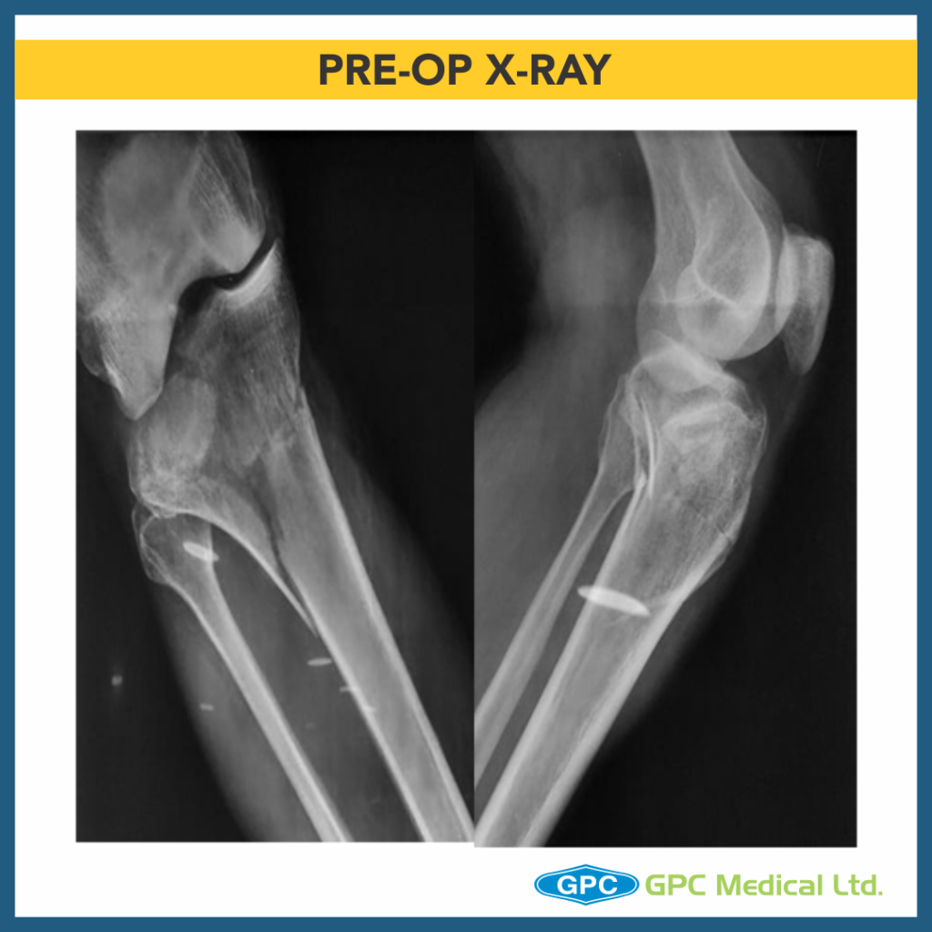
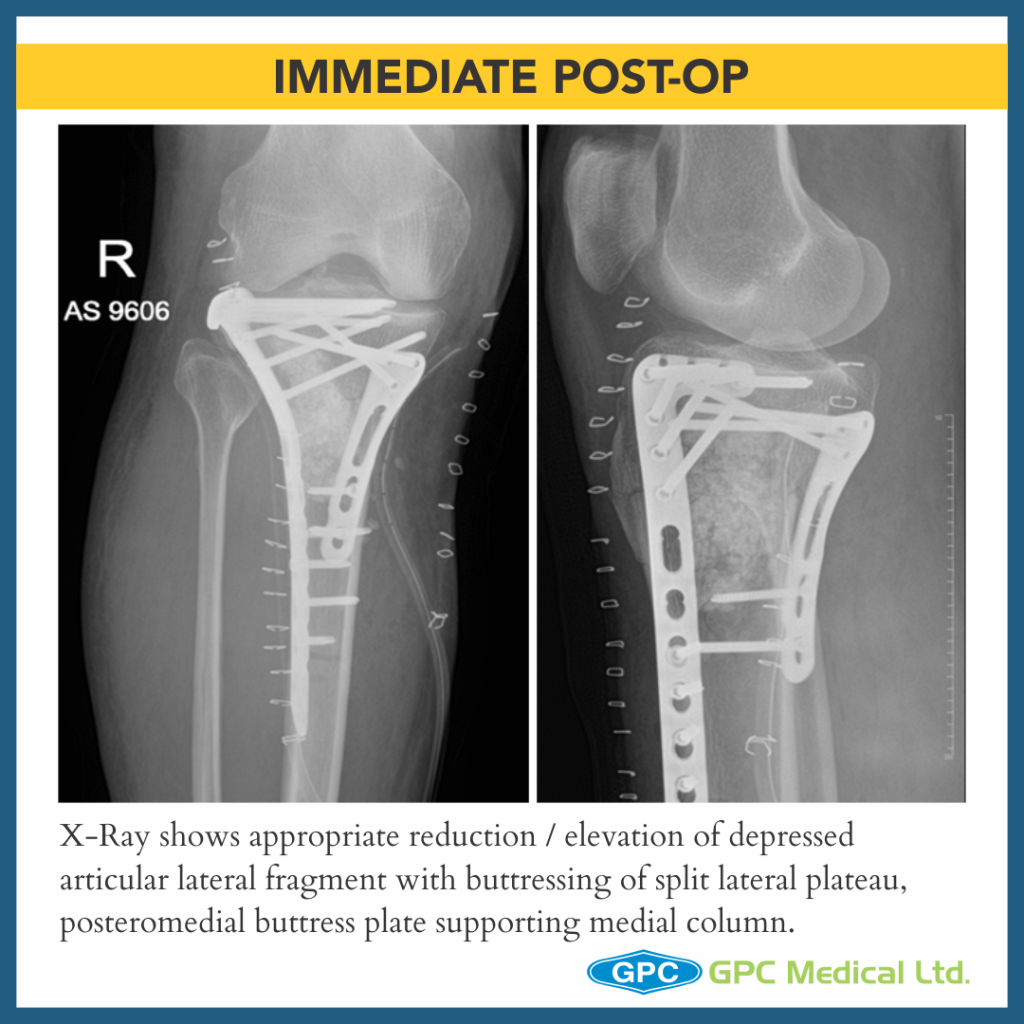
– X-Ray Right knee – Anteroposterior and Lateral Views

Diagnosis:
– Split Depression Lateral Tibial Plateau Fractures (Schatzkers Type 2).

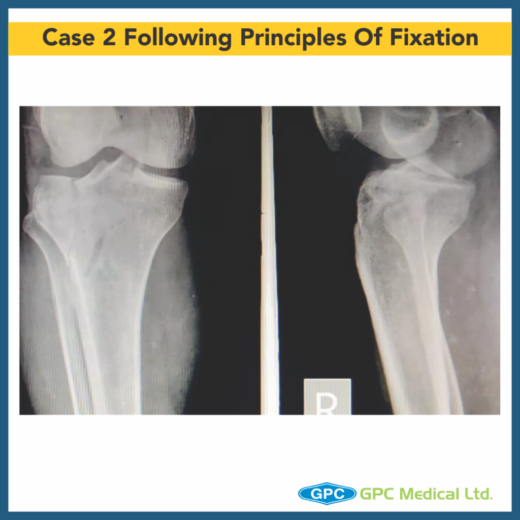
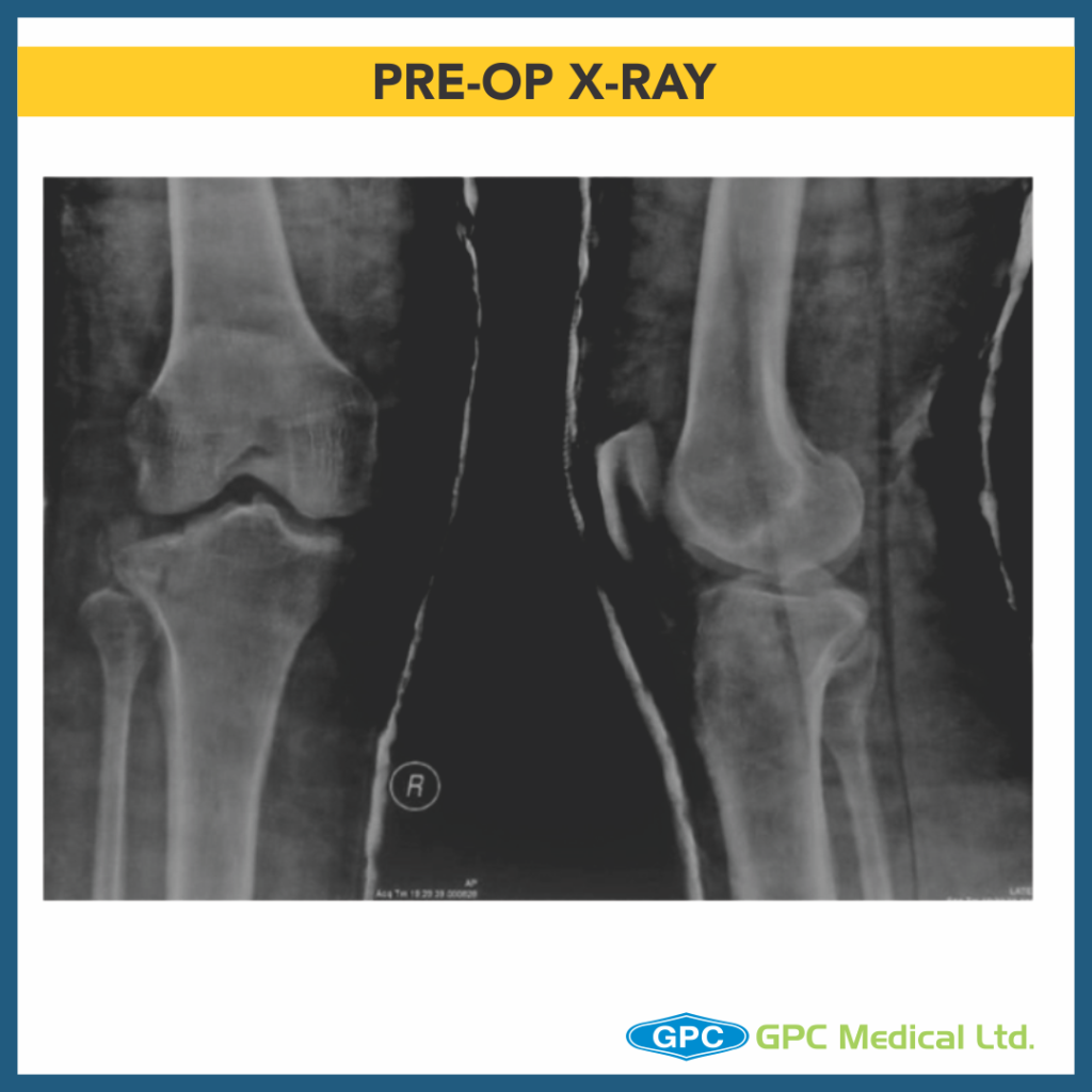
CASE 2

Clinical Presentation:
– 59 years Male.
– Alleged history of Road traffic accident.
– Severe pain in right knee with restricted knee ROM and inability to weight bear on right leg.

Radiological investigation:
– X-Ray Right knee – Anteroposterior and Lateral Views

Diagnosis:
– Medial Plateau Fracture With Split Depression Lateral Tibial Plateau Fractures With Meta-diaphyseal Dissociation (Schatzkers Type 6).

CASE 3

Clinical Presentation:
– 44 years Male.
– Alleged history of Road traffic accident.
– Severe pain in left knee with restricted knee ROM and inability to weight bear on left leg.

Radiological investigation:
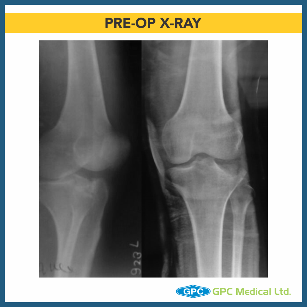
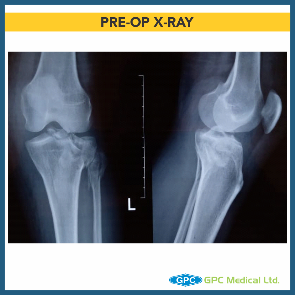
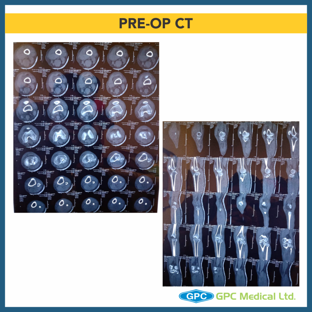
– X-Ray Left knee – Anteroposterior and Lateral Views

Diagnosis:
– Split Depression Lateral Tibial Plateau Fractures (Schatzkers Type 2).
CASE 4

Clinical Presentation:
– 18 years Female.
– Alleged history of Road traffic accident.
– Severe pain in left knee with restricted knee ROM and inability to weight bear on left leg.

Radiological investigation:
– X-ray Left knee – Anteroposterior and Lateral Views

Diagnosis:
– Depression Type Lateral Tibial Plateau Fractures (Schatzkers Type 3).

CASE 5

Clinical Presentation:
– 29 years Male.
– Alleged history of Road traffic accident.
– Severe pain in right knee with restricted knee ROM and inability to weight bear on right leg.

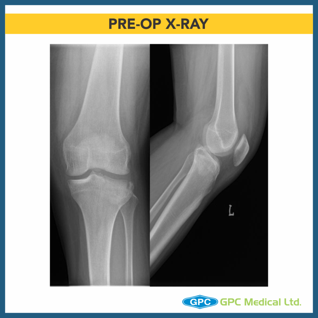
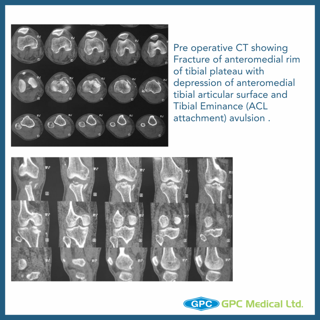
Radiological investigation:
– X-Ray Right knee – Anteroposterior and Lateral Views

Diagnosis:
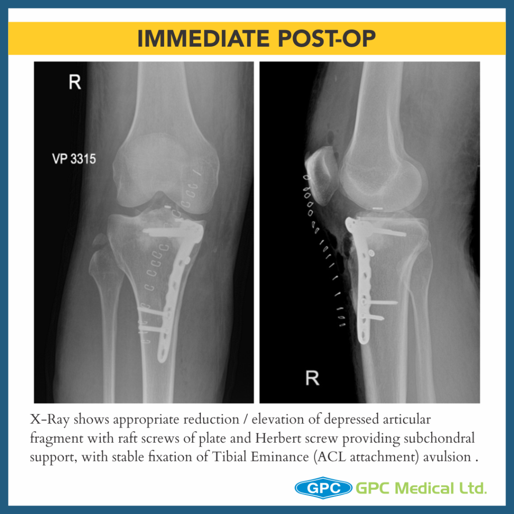
– Split Depression Type Anteromedial Tibial Plateau Fractures with Tibial Eminance (ACL attachment) avulsion.

Surgical Principles and Lacunae in Management

– Principles

  • Management for Periarticular Fractures is based on concept of Anatomical reduction of fracture.
  • To allow for Primary Bone Healing to happen, Absolute stability and Rigid fixation must be attained.
  • Use reduction technique that respects the biological principles of fixation (closed, minimally invasive or open).
  • Long term predictor of satisfactory outcome post surgery is restoration of articular suface and mechanical alignment restoration of limb.
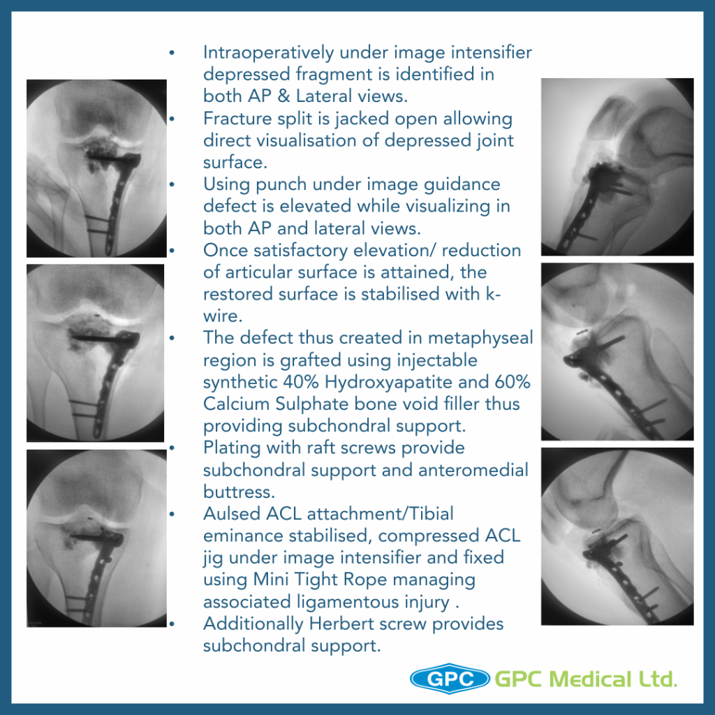
  • Once satisfactory elevation/ reduction of articular surface is attained, the restored surface is stabilised with k-wire.
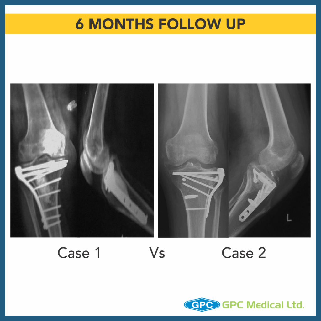
  • The defect thus created in metaphyseal region is grafted using either Natural bone graft (Cortcocancellous bone) or Synthetic bone void filler (injectable or granules) thus providing excellent subchondral support.
  • These Grafts remodel into host bone within 6-12 months.
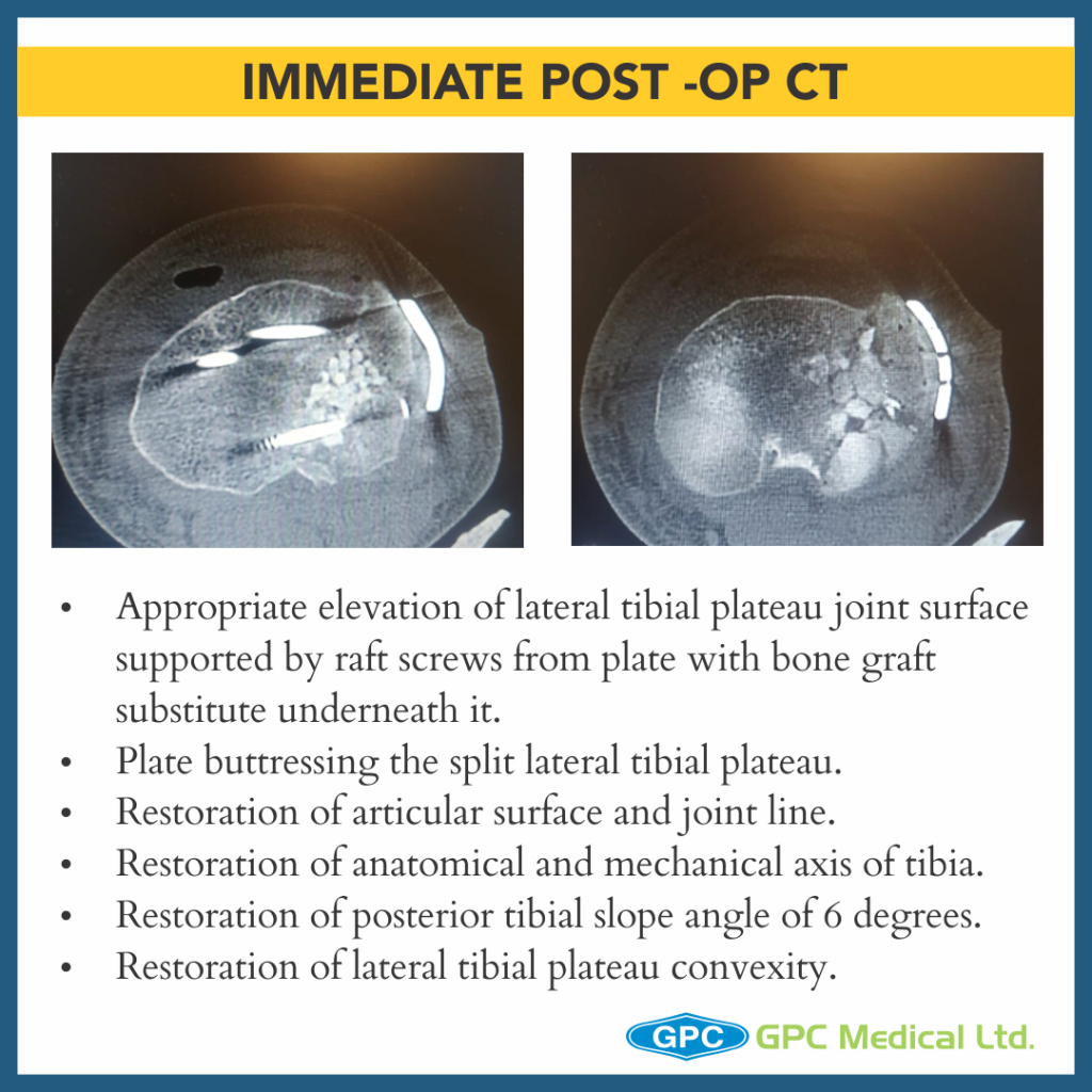
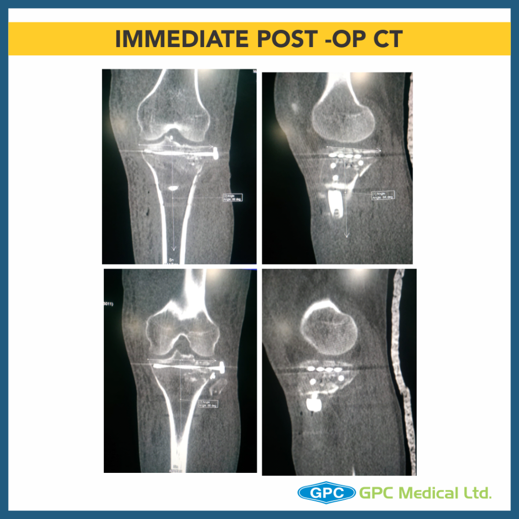
  • Finally raft screws of plate provide subchondral support and plate help in maintaining the long mechanical axis of limb.

– Lacunae

  • Failure to restore articular surface anatomically may result in long term post traumatic arthritis.
  • Medial tibial plateau is less forgiving as far as late consequences of a fracture because of smaller meniscus covering joint surface compared to lateral tibial plateau.

Proximal Tibia Fracture: Case Discussion

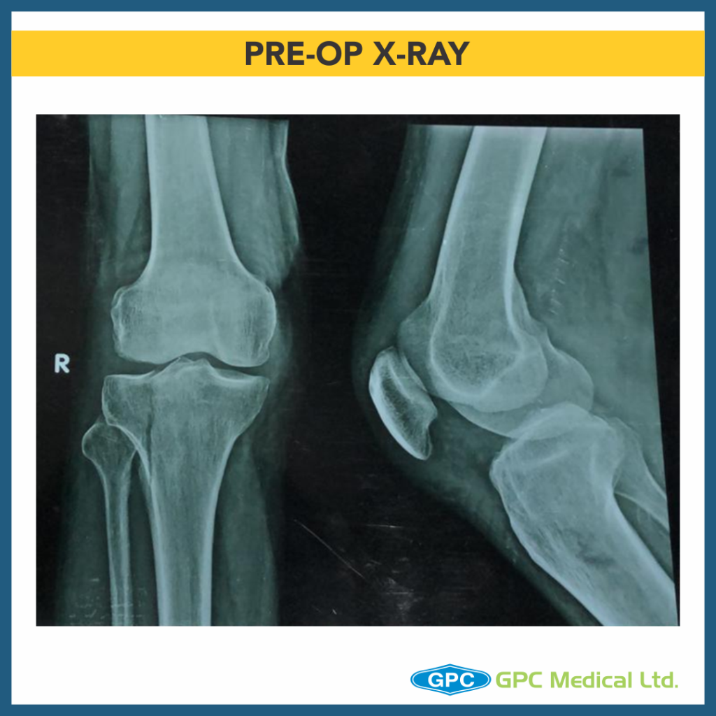
Today, we’re discussing proximal tibia fracture case. Pre-planning is the key! Sometimes for adequate visualisation of the articular surface, surgeons may prefer Arthroscopy, however a few surgeons prefer submeniscal arthrotomy for direct visualisation of fracture reduction.

Clinical History

  • 46 year old male
  • History of RTA
  • Presented on the day of Injury
  • Pain and swelling over right leg
  • Co-morbidities – nil

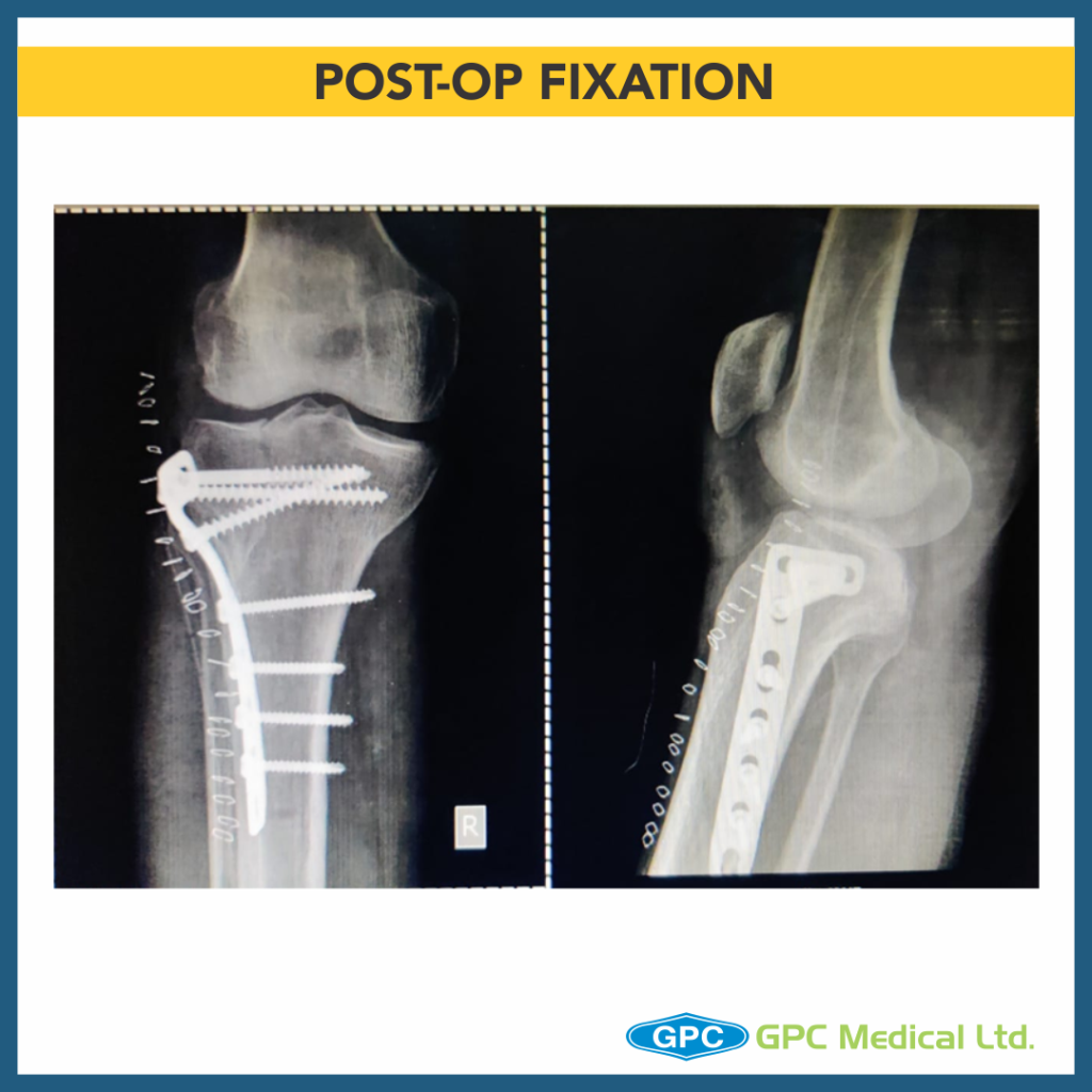
What are the issues with this fixation?

  • Fracture appears to be Split fracture of lateral tibial condyle (Schatzker Type 1).
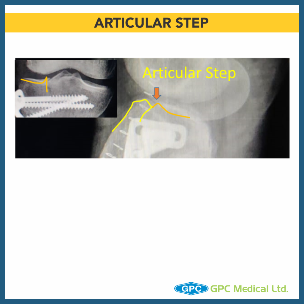
  • Articular step has not been corrected.
  • Compression not achieved at fracture site.
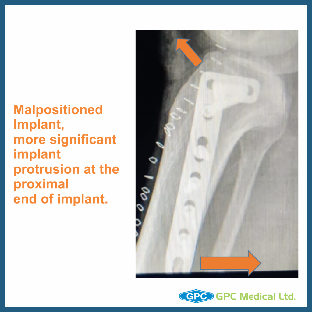
  • Hockey plate gives option of placement of only two screws at subchondral region, additional screws (outside plate) could have been used.
  • Plate is not positioned properly can lead to implant prominence.
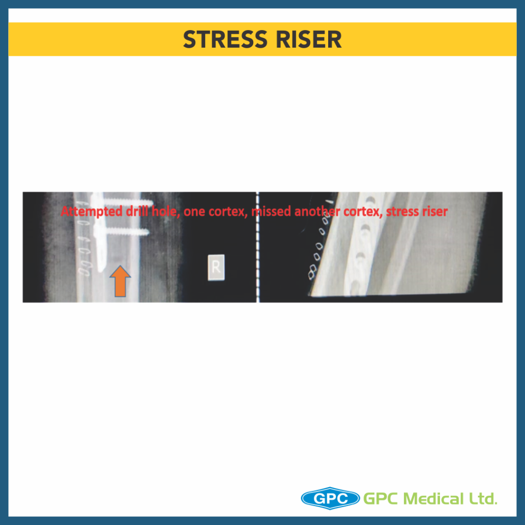
  • Stress riser at plate end due to attempted bone drill.

Which factors led to these issues during fixation?

  • Adequate exposure and visualization of the articular surface is of prime importance in fixation of such fractures.
  • These fractures require pre-surgical planning in terms of approach required for accessing each fracture with adequate visualization of articular surface.
  • Some Surgeons advocate use of arthroscopy to visualize the articular continuity/reduction whereas other group prefer submeniscal arthrotomy for direct visualization of fracture reduction.
  • These fractures appear simple and one tends to have minimum visualization of lateral fluoroscopy view in a hurry of fracture fixation.

How to troubleshoot these factors?

  • Pre-plan the surgery in-terms of fixation system- Internal fixation/ external fixation.(read implant manual for ideal site of placement of plates)
  • Plan the necessary approach.
  • Plan for bone grafting if articular depression is observed intraoperatively.
  • Always temporarily fix the reduction with K wires/ screw to prevent sagging of articular fragment and Visualize the articular congruity.
  • Confirm the fracture reduction in two planes.
  • Use of magnified fluoroscopy view of articular surface in anteroposterior and lateral view.

Author approach

  • Adequate visualization of the fracture with submeniscal arthrotomy.
  • Reduction of fracture and compression is temporarily achieved with condylar compression clamps and temporary fixation with K wires.
  • Countersunked Lag screw at the apex of the fracture fragment.
  • T shaped plate instead of L shaped Plate as fracture line is in anterior one third tibia(lateral view)
  • Longer plate for spacing out screws.
  • Backup plan:
    • Additional lag screw in condylar region(prior to plate fixation)
    • Window on contralateral cortex for elevating articular surface

Tibial Plateau Fractures: Depressed Fragment Elevation Technique

Today, we’re going to talk about Tibial plateau fractures & the depressed fragment elevation technique! Anatomic reduction of the fracture & rigid fixation is the key!

CASE 1

Clinical Presentation:

  • 60 years female.
  • Slip and fall at home.
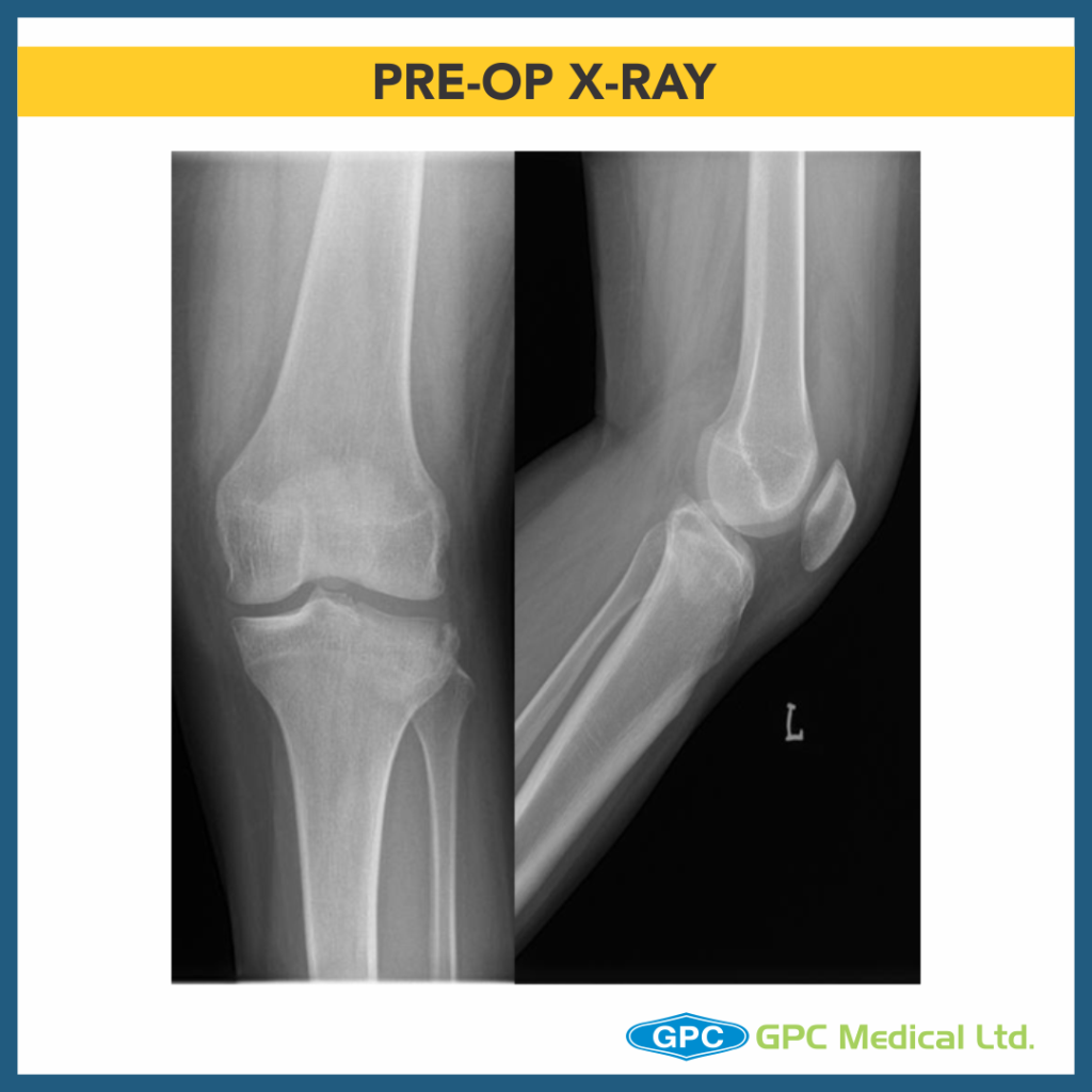
  • Severe pain in right knee with restricted knee ROM and inability to weight bear on right leg.

Radiological Investigation:

  • X-Ray Right knee – Anteroposterior and Lateral Views

Diagnosis:

  • Split Depression Lateral Tibial Plateau Fractures (Schatzkers Type 2).
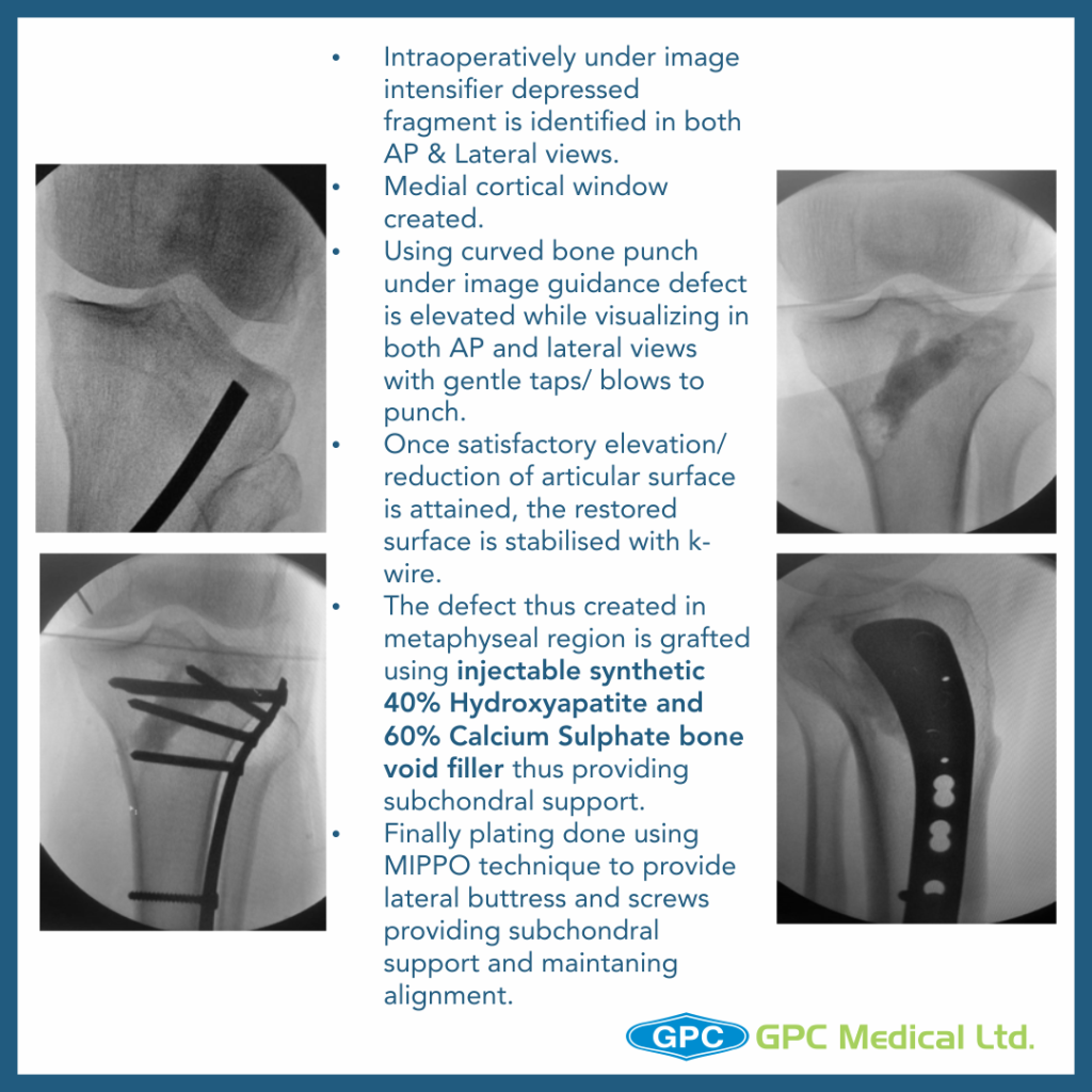
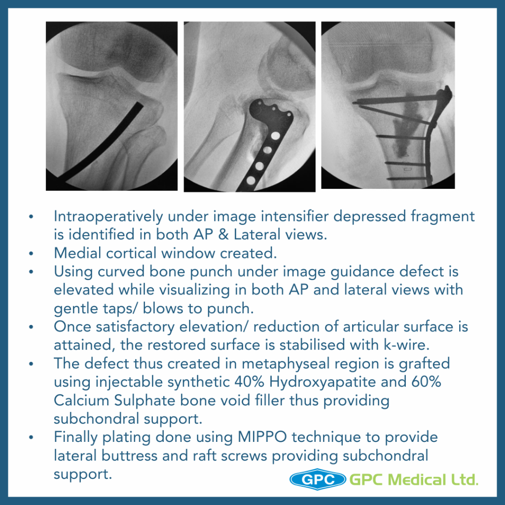
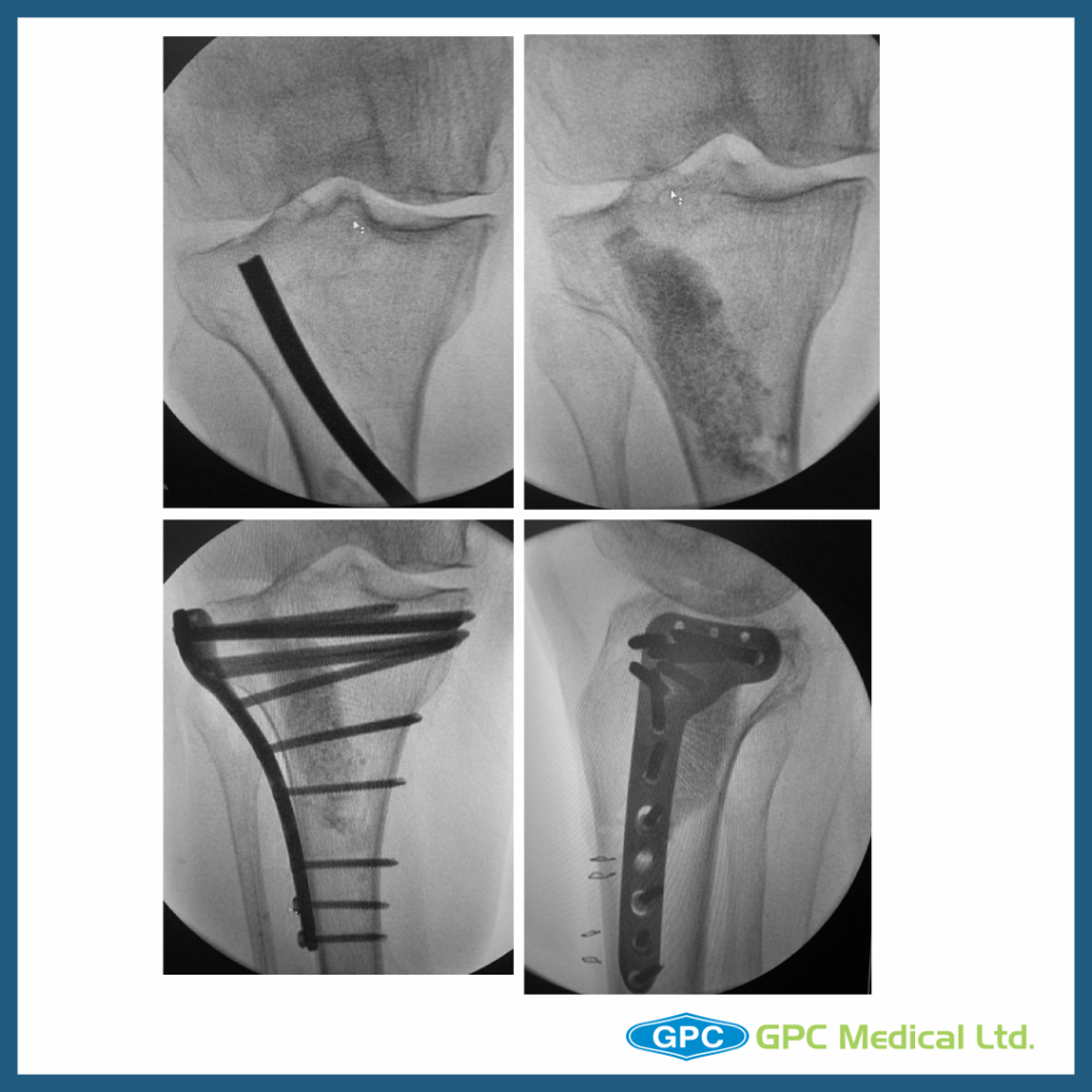
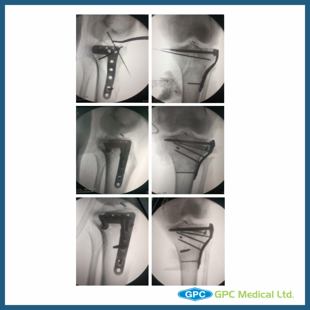
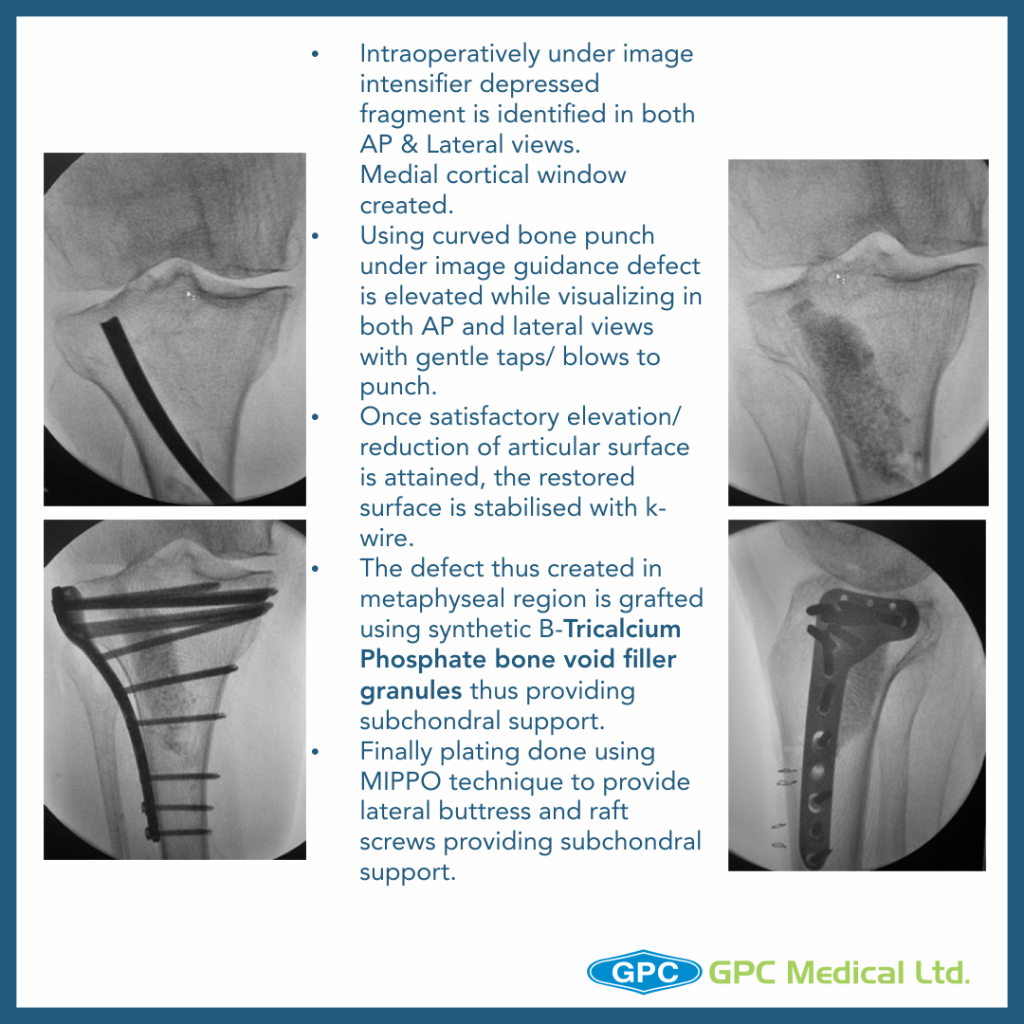
  • Intraoperatively under image intensifier depressed fragment is identified in both AP & Lateral views.
  • Medial cortical window created.
  • Using curved bone punch under image guidance defect is elevated while visualizing in both AP and lateral views with gentle taps/ blows to punch.
  • Once satisfactory elevation/ reduction of articular surface is attained, the restored surface is stabilised with k-wire.
  • The defect thus created in metaphyseal region is grafted thus providing subchondral support.
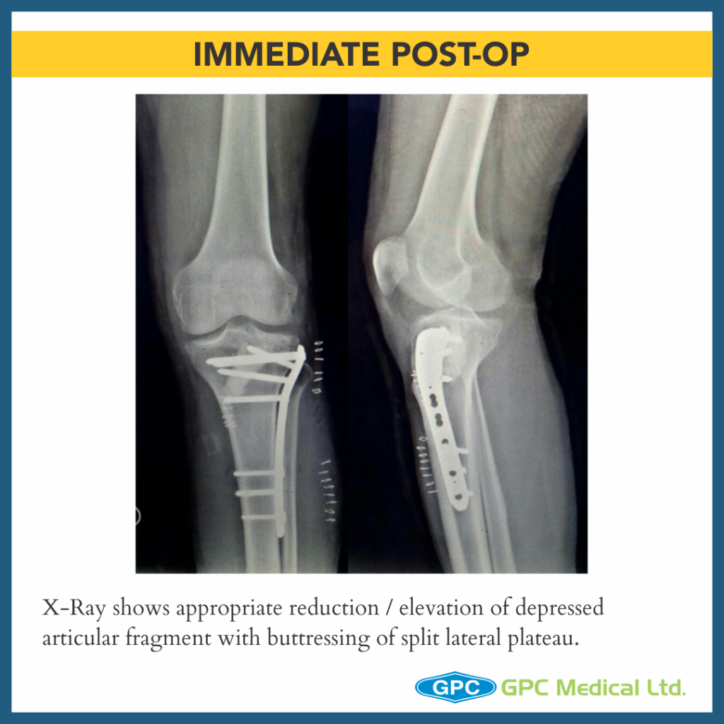
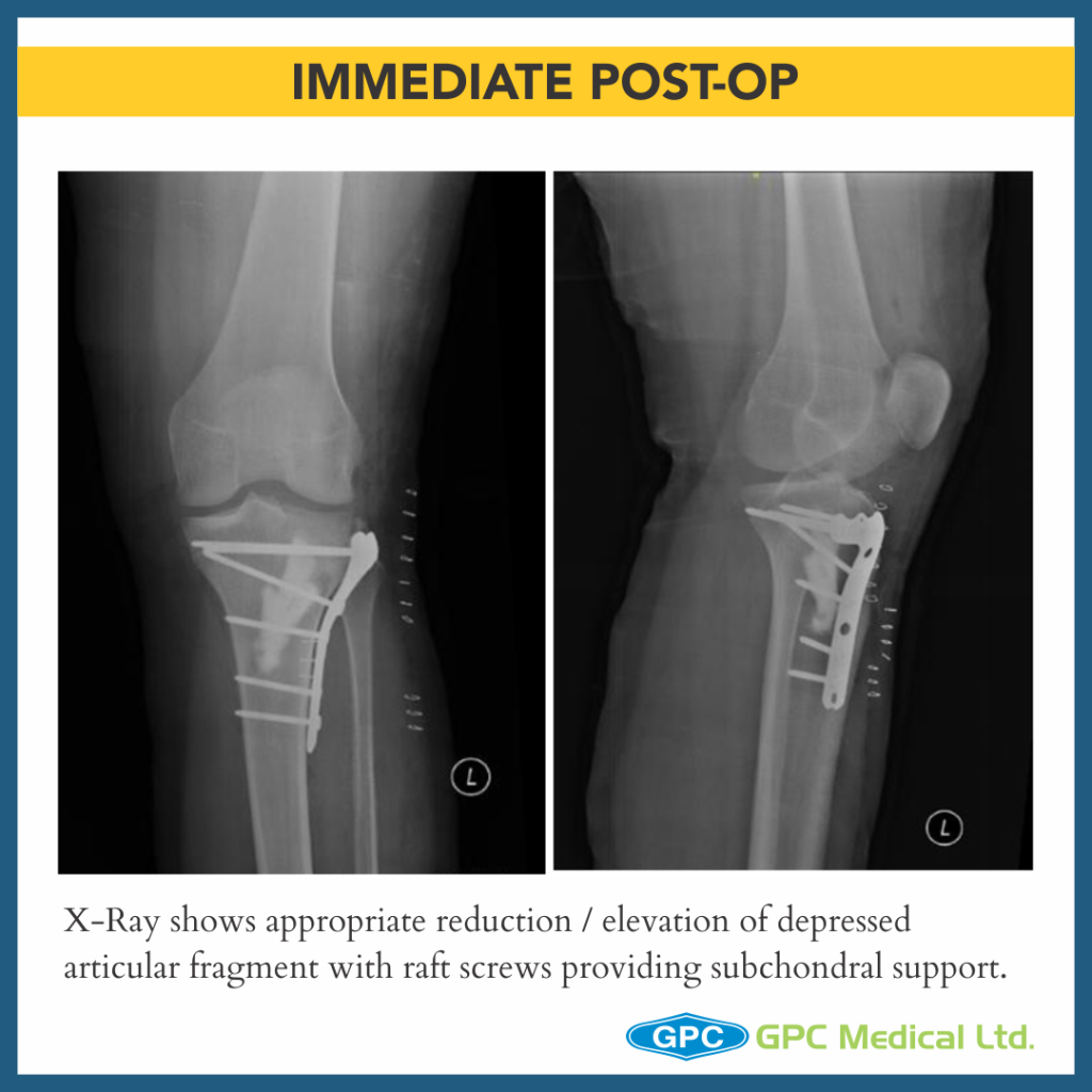
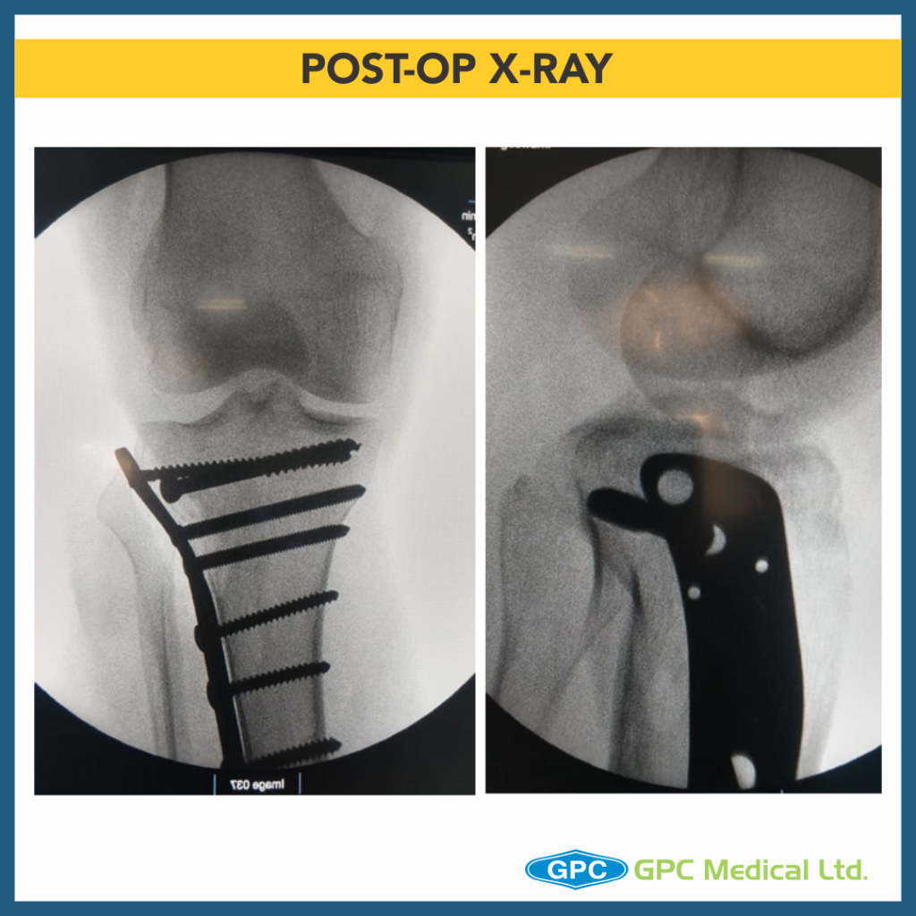
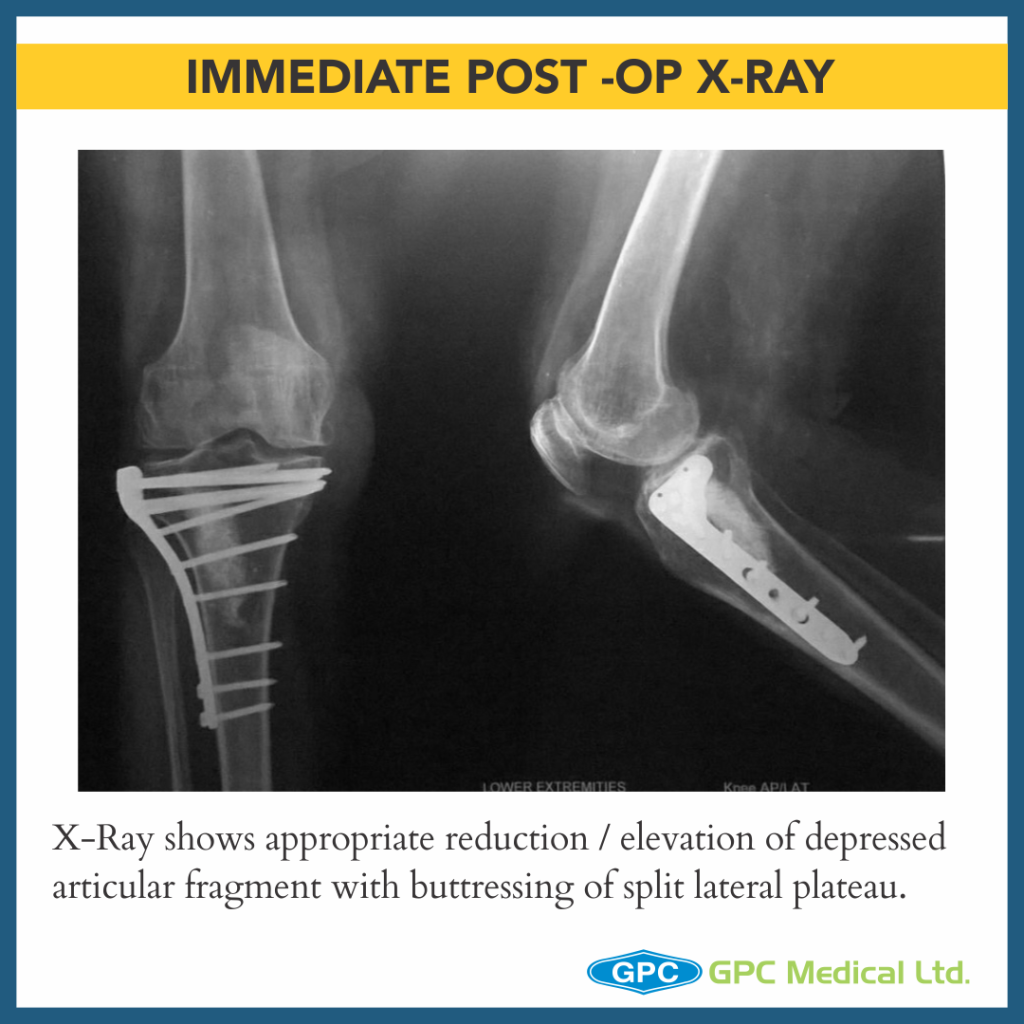
  • Finally plating done using MIPPO technique to provide lateral buttress and raft screws providing subchondral support.
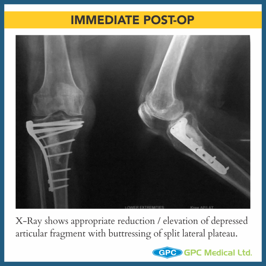
X-Ray shows appropriate reduction / elevation of depressed articular fragment with buttressing of split lateral plateau.

CASE 2

Clinical Presentation:

  • 39 years male.
  • Motor Vehicle Accident.
  • Severe pain in left knee with restricted knee ROM and inability to weight bear on left leg.

Radiological Investigation:

  • X-Ray Left knee – Anteroposterior and Lateral Views

Diagnosis:

  • Split Depression Fracture Lateral Tibial Plateau with Tibial Eminance (ACL attachment) avulsion and MCL Injury
  • Intraoperatively under image intensifier depressed fragment is identified in both AP & Lateral views.
  • Fracture split is jacked open allowing direct visualisation of depressed joint surface.
  • Using punch (here a curette) under image guidance defect is elevated while visualizing in both AP and lateral views.
  • Once satisfactory elevation/ reduction of articular surface is attained, the restored surface is stabilised with k-wire.
  • The defect thus created in metaphyseal region is grafted thus providing subchondral support.
  • Split repositioned, compressed and stabilised with K-wires.
  • Finally plating done using MIPPO technique to provide lateral buttress and raft screws providing subchondral support.
  • Aulsed ACL attachment/Tibial eminance stabilised, compressed ACL jig under image intensifier and fixed using Mini Tight Rope
  • Split posterolateral fragment reduced, stabilised and fixed with 4mm CCS in compression mode, restoring congurity of lateral tibial plateau.
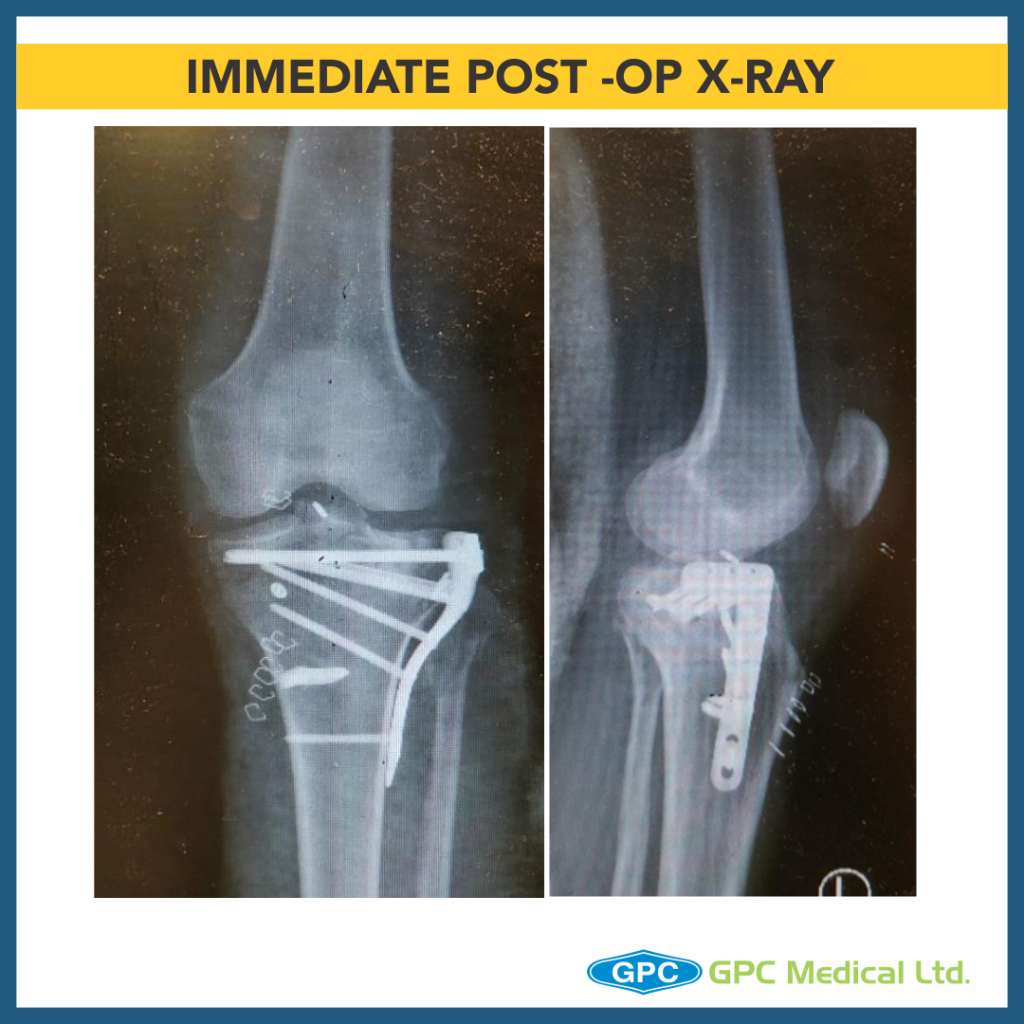
  • MCL was intraoperatively found to be avulsed from its tibial attachment site, reenforced with ethibond and fixed using bone staple.
  • Thus, managing fracture with associated multi-ligamentous injury in toto.

Surgical Principles and Lacunae in Management

  • Principles
    • Management for Periarticular Fractures is based on concept of Anatomical reduction of fracture.
    • To allow for Primary Bone Healing to happen, Absolute stability and Rigid fixation must be attained.
    • Use reduction technique that respects the biological principles of fixation (closed, minimally invasive or open).
  • Lacunae
    • Failure to restore articular surface anatomically may result in long term post traumatic arthritis.