Hip Fractures Fixation with Nails – Everything you need to know

According to a survey, the number of hip fractures is expected to surpass 6 million worldwide by the year 2050. Hip fractures are breaks in the upper portion of the femur that usually occurs in elderly patients whose bones have become weakened by osteoporosis. In case of younger patients, hip fractures occur due to a high-energy event, such as a fall from a ladder or vehicle collision. Most of these fractures occur in older patients who are injured in household or community falls.

Hip fractures tend to be very painful. This is why prompt surgical treatment is recommended. Treating the fracture and getting the patient out of bed as soon as possible helps prevent medical complications later on such as bed sores, blood clots and pneumonia. Disorientation can also occur in very old patients due to prolonged bed rest which makes rehabilitation and recovery much more difficult.

Orthopedic implants like nails play an important role in the fixation process. The choice of the implant material is very crucial as it influences rigidity, corrosion, bio-compatibility and tissue receptivity. The surface morphology of the implants also affects its stability within the skeleton or the surrounding cement mantle.

GPC Medical has been serving the medical world as a leading manufacturer of medical equipment and healthcare systems for more than 20 years. As one of the top manufacturers and suppliers of medical equipment and affordable healthcare solutions in India, GPC Medical strives continuously to innovate medical solutions that enriches the lives of people. We are an ISO 9001, ISO 13485 certified company that exports and supplies medical equipment and surgical instruments worldwide.

GPC Medical manufactures a wide range of nails for hip fractures that are made from the best raw materials with ergonomic design. Let’s take a look at the various types of hip nails:

intraHEAL Proximal Hip Stabilizing Nail (ILBS59)

Our IntraHEAL Proximal Hip Stabilizing Nail is the best-in-class nail in the market for intertrochanteric fractures of femur. Our IntraHEAL Proximal Hip Stabilizing Nail is US FDA approved and designed to provide superior bio-mechanical intramedullary stabilization. In stable fractures, it provides circumferential compression at the fracture site and transfers axial load to the bone.

Advanced Features:

  • Our Nails are equipped with a self-tapping lag screw for easy insertion.
  • Available in length ranging from 180-220mm, and proximal nail angle of 130°.
  • It has a cannulated nail for guide wire controlled insertion.
  • It also has a set screw that inhibits rotation of the proximal lag screw & simultaneously allows sliding of the lag screw.
  • It has a single distal locking option to prevent rotation in complex fracture.
  • Universal nail for the right & left hip.
  • Available in stainless steel and titanium.

intraHEAL Proximal Femoral Nail, Advanced (PFA09-12)

Our second nail in the series, IntraHEAL Proximal Femoral Nail is an advanced nail implant ideal for the treatment of Pertrochanteric fractures, Intertrochanteric fractures & high subtrochanteric fractures. An ideal implant for the treatment of unstable fractures, the nail is ergonomically designed so that it can be easily inserted and is especially useful for elderly persons with osteoporosis. and is further equipped with a cannulated blade to provide enhanced angular and rotation stability.

Advanced Features:

  • Anatomically designed for optimal fit in the femur.
  • The nail comes with a signature design cannulated blade that provides increased stability and helps compression of the cancellous bone while also providing angular and rotational stability.
  • The nail also allows early weight bearing and mobilization.
  • Both nail and blade are also cannulated.
  • Available in titanium only.

intraHEAL Proximal Hip Stabilizing Nail 3 (PHN3)

Our 3rd nail for hip fractures, intraHEAL Proximal Hip Stabilizing Nail 3 (PHN3) is an advanced version of Proximal hip stabilizing nail that is specifically designed for Asian population. The nail is used in intertrochanteric fractures, high subtrochanteric fractures and per subtrochanteric fractures. The nail has been ergonomically designed for minimally invasive surgery and conforms to international quality standards.

Advanced Features:

  • The proximal diameter is 15.5 mm, to minimize the incision length required for minimally invasive surgery.
  • Available in three neck angles- 120, 125, 130 degrees to accommodate various anatomies.
  • Cannulated nail for guide wired controlled insertion.
  • The thread design of the lag screw enables superior cut out strength from the cancellous bone.
  • Short nail has one distal locking screw & the long nail has two.

Contact us to know more about our nails. We at GPC Medical are always welcome to any queries regarding our products.

Inter-Trochanteric Fractures Series – PART 10

Significance with regards to Lateral Wall – Part II

We are here with another session of Inter-Trochanteric Fractures. This is the second part of our video on the significance of the lateral wall. So, watch the video to know more.

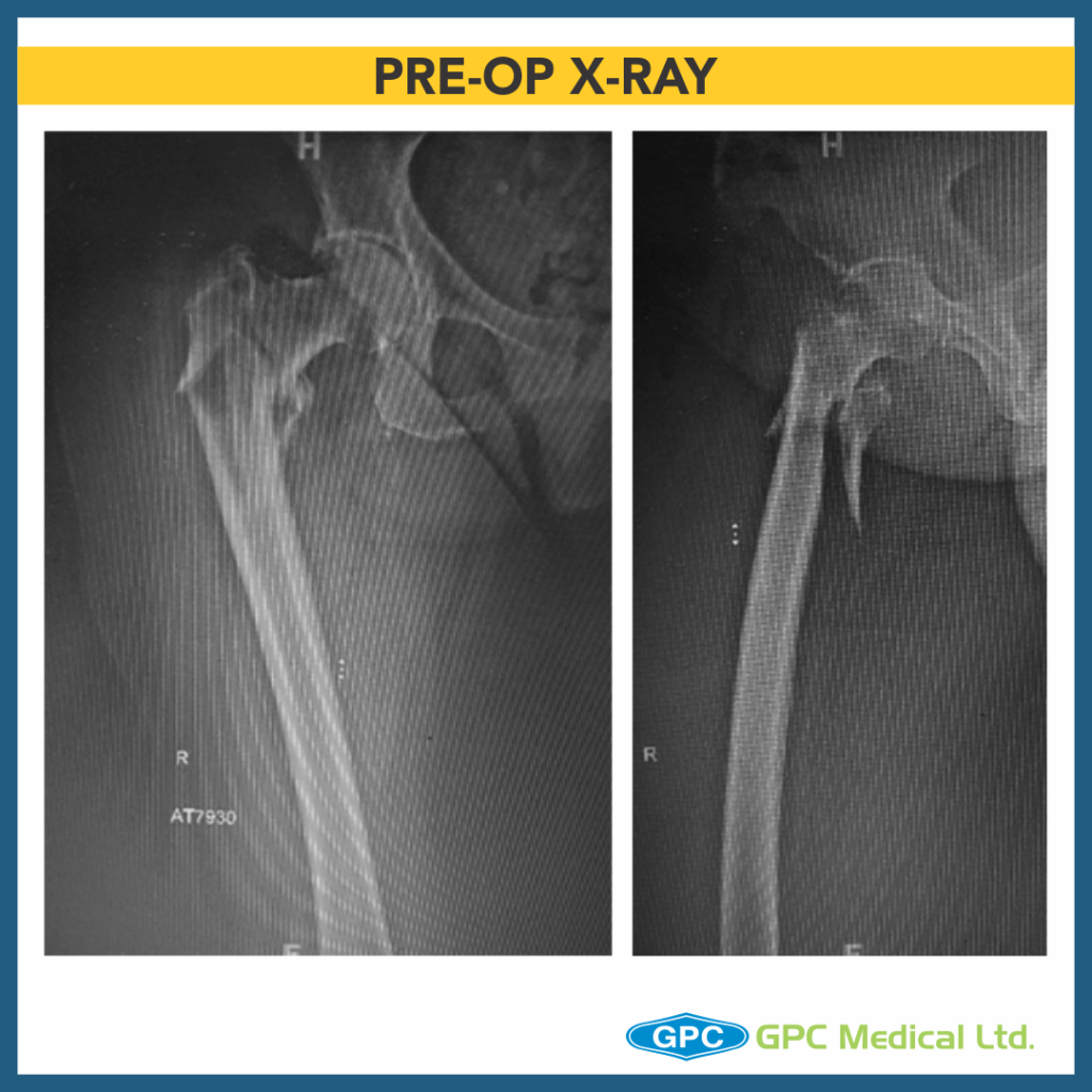
CASE 1

Clinical Presentation:
– 80 years Male.
– Fall at home.
– Severe pain in right hip with inability to stand

Radiological investigation:
– X-Ray pelvis with both hips –Anteroposterior and lateral view

Diagnosis:
– Unstable intertrochanteric fracture with subtrochanteric extension.

CASE 2

Clinical Presentation:
– 84 years female.
– Fall in bathroom.
– Severe pain in right hip with inability to stand

Radiological investigation:
– X-Ray pelvis with both hips –Anteroposterior view

Diagnosis:
– Unstable intertrochanteric fracture with subtrochanteric extension.

Surgical Principles and Lacunae in Management

  • Standard of management for unstable intertrochanteric fracture is proximal femoral nailing because of its superior biomechanical properties.
  • Lacunae
    • Post operative and long term stability with acceptable outcome needs maintenance of medial calcar in AP radiograph and anterior calcar in lateral radiograph.
    • Lateral trochanteric wall has been recognized as an important predictor of stability in intertrochanteric fractures.
    • PFN(intramedullary nails) because it bypasses the lateral wall and acts as a prosthetic lateral cortex medial to broken/thinned out lateral wall gives better biomechanical advantage with lesser lateral migration of the proximal fragment as it abuts the medial aspect of nail.
    • Also, nail by similar mechanism prevents limb shortening thus helping maintaining limb length.
    • There has been lot of research concerning lateral wall integrity/reconstruction using additional plate, wiring with regards to functional outcome with regards to abductor muscle function by still no discrete recommendations have been made. (as in our cases though fracture union was good in both cases, in case 2 where lateral wall was stabilised with additional SS wiring long term lateral wall displacement is less compared to case 1).

Inter-Trochanteric Fractures Series – PART 9

Significance with regards to Lateral Wall – Part I

Today is the part 9 of the Inter-Trochanteric fractures, where we’re going to talk about the significance of the lateral wall. Lateral trochanteric wall has been recognised as an important predictor of the stability in intertrochanteric fractures.

CASE

Clinical Presentation:
– 75 years female.
– Slip and fall at home.
– Severe pain in left hip with inability to stand

Radiological investigation:
– X-Ray pelvis with both hips – Anteroposterior view

Diagnosis:
– Unstable intertrochanteric fracture.

Surgical Principles and Lacunae in Management

  • Standard of management for unstable intertrochanteric fracture is proximal femoral nailing because of its superior biomechanical properties.
  • Lacunae
    • Post operative and long term stability with acceptable outcome needs maintenance of medial calcar in AP radiograph and anterior calcar in lateral radiograph.
    • It can be obtained by proper patient positioning and closed manipulation or minimally invasive technique (like using Homman lever through nail entry point incision as in this case)
    • Lateral trochanteric wall has been recognized as an important predictor of stability in intertrochanteric fractures.
    • PFN (intramedullary nails) because it bypasses the lateral wall and acts as a prosthetic lateral cortex medial to broken/thinned out lateral wall gives better biomechanical advantage.
    • Also, PFN (intramedullary nails) prevent excessive collapse of fracture by providing lateral buttress and prevent medial displacement of femoral shaft (as seen in this case after implant removal and fracture healing).

Inter-Trochanteric Fractures Series – PART 8

Continuing with the IT fracture series, we’re going to talk about significance of nail length today as well. Choosing the optimal nail length is critical in any fracture management.

CASE 1

Clinical Presentation:
– 84 years Male.
– Fall at home.
– Severe pain in left hip with inability to stand

Radiological investigation:
– X-Ray pelvis with both hips –Anteroposterior and lateral view

Diagnosis:
– Unstable intertrochanteric fractutre with subtrochanteric extension.

CASE 2

Clinical Presentation:
– 84 years female.
– Fall in bathroom.
– Severe pain in right hip with inability to stand

Radiological investigation:
– X-Ray pelvis with both hips –Anteroposterior view

Diagnosis:
– Unstable intertrochanteric fractutre with subtrochanteric extension.

Surgical Principles and Lacunae in Management

  • Standard of management for unstable intertrochanteric fracture with subtrochanteric extension is long proximal femoral nailing because of its superior biomechanical properties.
  • Lacunae
    • In Case 1 where intraoperative reduction was satisfactory but long nail caused impingement on distal anterior femoral cortex, which may lead to anterior thigh pain and rarely even a supracondylar fracture.
  • In Case 2 where intraoperative reduction was satisfactory and long nail tip is in centre of distal femur medullary canal not causing any impingement on distal anterior femoral cortex is the ideal position.
  • Though, attaining central position distally is desirable but is not always possible because of mismatch in femoral nail radius of curvature and native femur anatomy.

Inter-Trochanteric Fractures Series – PART 7

In continuation to our Inter-Trochanteric Fractures series, we’re going to talk about the significance of nail length in IT fractures.

CASE 1

Clinical Presentation:
– 79 years female.
– Fall at home.
– Severe pain in left hip with inability to stand

Radiological investigation:
X-Ray pelvis with both hips –Anteroposterior view

Diagnosis:
Unstable intertrochanteric fractutre.

CASE 2

Clinical Presentation:
84 years female.
Fall in bathroom.
Severe pain in right hip with inability to stand

Radiological investigation:
X-Ray pelvis with both hips –Anteroposterior and Lateral view

Diagnosis:
Unstable intertrochanteric fractutre.

Surgical Principles and Lacunae in Management

  • Standard of management for unstable intertrochanteric fracture is proximal femoral nailing because of its superior biomechanical properties.
  • Lacunae
    • In cases where if shorter (170mm,200mm) nails are used in wider canal there are chances of movement of nail within the femoral canal inturn leading to loss of fracture reduction.
  • Mid length (240mm) nails can prevent the wiper motion of distal stem because spans the entire length of isthmus.
  • Though, there is no ideal nail length as it depends on fracture configuration and femoral canal anatomy, nail length of 240mm tends to provide most satisfactory outcomes.

Inter-Trochanteric Fractures Series – PART 6

Inter-Trochanteric Fractures Series continues… Today, we’re going to talk about significance of position of lag screw in proximal femur nailing.

CASE 1

Clinical Presentation:
– 87 years female.
– Fall in bathroom.
– Severe pain in right hip with inability to stand

Radiological investigation:
X-Ray pelvis with both hips – Anteroposterior view

Diagnosis:
Unstable intertrochanteric fractutre.

CASE 2

Clinical Presentation:
– 78 years female.
– Slip and fall at home.
– Severe pain in left hip with inability to stand

Radiological investigation:
X-Ray pelvis with both hips – Anteroposterior view

Diagnosis:
Unstable intertrochanteric fractutre.

Surgical Principles and Lacunae in Management

  • Standard of management for unstable intertrochanteric fracture is proximal femoral nailing because of its superior biomechanical properties.
  • Lacunae
    • Post operative and long term stability with acceptable outcome needs maintenance of medial calcar in AP and anterior calcar in lateral radiograph.
    • Carry out open reduction if result is unsatisfactory.
  • Though fracture reduction may be satisfactory but eccentric placement of lag screw leads to loosening at bone- lag screw interface with resultant pain and failure and should be avoided.
  • Ideal lag screw position is centre-centre in both AP and lateral views.
  • If it is in unacceptable position inferior-posterior in AP and lateral views is better than superior-anterior position(undesirable position with high chances of lag screw cut out).

Inter-Trochanteric Fractures Series – PART 5

In continuation to our IT Fracture series, we’re going to talk about significance of locking lag screws in proximal femur nailing.

CASE 1

Clinical Presentation:
– 88 years female.
– Slip and fall at home.
– Severe pain in left hip with inability to stand

Radiological investigation:
– X-Ray pelvis with both hips – Anteroposterior view
– X-Ray left hips – Lateral view

Diagnosis:
– Unstable intertrochanteric fracture.

CASE 2

Clinical Presentation:
– 84 years female.
– Fall on wet floor at home.
– Severe pain in left hip with inability to stand

Radiological investigation:
– X-Ray pelvis with both hips – Anteroposterior view
– X-Ray left hips – Lateral view

Diagnosis:
– Unstable intertrochanteric fracture.

Surgical Principles and Lacunae in Management

  • Standard of management for unstable intertrochanteric fracture is proximal femoral nailing because of its superior biomechanical properties.
    • Tip apex distance should be kept <25 mm to obtain maximum purchase in subchondral bone.
    • Centre-Centre position of lag screw in both AP and Lateral view is the most desirable.
  • Lacunae
    • In osteoporotic bones because of poor bone quality there is less secure hold of lag screw in bone, with increased chances of screw backout.
    • Locking the lag screw maintains proximal femoral anatomy, minimizes femoral neck shortening and prevents screw backout.
    • Locking the lag screw maintains proximal femoral anatomy, minimizes femoral neck shortening and prevents screw backout.

Inter-Trochanteric Fractures Series – PART 4

As part of our ongoing series of treatment of IT fractures, we are continuing the discussion on how important it is to maintain the anterior calcar for a better prognosis.

CASE 1

Clinical Presentation:
– 78 years male.
– Fall in bathroom.
– Severe pain in right hip with inability to stand

Radiological investigation:
X-Ray right hip – Anteroposterior view

Diagnosis:
Unstable intertrochanteric fractutre.

CASE 2

Clinical Presentation:
– 91 years female.
– Slip and fall at home.
– Severe pain in right hip with inability to stand

Radiological investigation:
– X-Ray pelvis with both hips – Anteroposterior and Lateral view

Diagnosis:
– Unstable intertrochanteric fractutre.

Surgical Principles and Lacunae in Management

  • Standard of management for unstable intertrochanteric fracture is proximal femoral nailing because of its superior biomechanical properties.
  • Lacunae
    • Post operative and long term stability with acceptable outcome needs maintenance of anterior calcar in lateral radiograph.
    • It can be obtained by proper patient positioning and closed manipulation or minimally invasive technique (like using Homman lever as in case 2 )
    • Carry out open reduction if result is unsatisfactory.
  • Quality of reduction has been classified into three types based on reduction of anterior calcar ( Type1: Reduced; Type2: Anterior displaced proximal fragment; Type 3: Posterior displaced proximal fragment)
  • Studies have shown that amount of sliding of proximal fragment is dependent on appropriate maintenance of anterior calcar with minimum sliding in Type 1 followed by Type 2 and finally Type 3 during controlled collapse of weight bearing and hence fracture union.

Inter-Trochanteric Fractures Series – PART 3

This week, we will continue to talk about Significance of Anterior calcar as part of our Intertrochanteric Fracture series.

CASE 1

PRE OP X-RAY

Clinical Presentation:
– 77 years female.
– Fall in drawing room.
– Severe pain in left hip with inability to stand

Radiological investigation:
– X-Ray pelvis with both hips – Anteroposterior view

Diagnosis:
– Unstable intertrochanteric fractutre.

CASE 2

PRE OP X-RAY

Clinical Presentation:
– 86 years female.
– Slip and fall at home.
– Severe pain in left hip with inability to stand

Radiological investigation:
– X-Ray pelvis with both hips – Anteroposterior view

Diagnosis:
– Unstable intertrochanteric fractutre.

Surgical Principles and lacunae in Management

  • Standard of management for unstable intertrochanteric fracture is proximal femoral nailing because of its superior biomechanical properties.
  • Lacunae
    • Post operative and long term stability with acceptable outcome needs maintenance of anterior calcar in lateral radiograph.
    • It can be obtained by proper patient positioning and closed manipulation or minimally invasive technique (like using Homman lever through nail entry point incision as in case 2 )
    • Carry out open reduction if result is unsatisfactory.
    • Quality of reduction has been classified into three types based on reduction of anterior calcar ( Type1: Reduced; Type2: Anterior displaced proximal fragment; Type 3: Posterior displaced proximal fragment)
    • Studies have shown that amount of sliding of proximal fragment is dependent on appropriate maintenance of anterior calcar with minimum sliding in Type 1 followed by Type 2 and finally Type 3 during controlled collapse of weight bearing and hence fracture union.

Inter-Trochanteric Fractures Series – PART 2

Today is the part 2 of the Inter-Trochanteric Fracture series. Here we will show the significance of maintaining anterior calcar.

CASE 1

PRE OP X-RAY

Clinical Presentation:
– 87 years female.
– Fall in bathroom.
– Severe pain in right hip with inability to stand

Radiological investigation:
– X-Ray pelvis with both hips – Anteroposterior view

Diagnosis:
– Unstable intertrochanteric fractutre.

CASE 2

PRE OP X-RAY

Clinical Presentation:
– 75 years female.
– Slip and fall at home.
– Severe pain in left hip with inability to stand

Radiological investigation:
– X-Ray pelvis with both hips – Anteroposterior view

Diagnosis:
– Unstable intertrochanteric fracture.

Surgical Principles and Lacunae in Management

  • Standard of management for unstable intertrochanteric fracture is proximal femoral nailing because of its superior biomechanical properties.
  • Lacunae
    • Post operative and long term stability with acceptable outcome needs maintenance of anterior calcar in lateral radiograph.
    • It can be obtained by proper patient positioning and closed manipulation or minimally invasive technique (like using Homman lever through nail entry point incision as in case 2 )
    • Carry out open reduction if result is unsatisfactory.