Hip Fractures Fixation with Nails – Everything you need to know

According to a survey, the number of hip fractures is expected to surpass 6 million worldwide by the year 2050. Hip fractures are breaks in the upper portion of the femur that usually occurs in elderly patients whose bones have become weakened by osteoporosis. In case of younger patients, hip fractures occur due to a high-energy event, such as a fall from a ladder or vehicle collision. Most of these fractures occur in older patients who are injured in household or community falls.

Hip fractures tend to be very painful. This is why prompt surgical treatment is recommended. Treating the fracture and getting the patient out of bed as soon as possible helps prevent medical complications later on such as bed sores, blood clots and pneumonia. Disorientation can also occur in very old patients due to prolonged bed rest which makes rehabilitation and recovery much more difficult.

Orthopedic implants like nails play an important role in the fixation process. The choice of the implant material is very crucial as it influences rigidity, corrosion, bio-compatibility and tissue receptivity. The surface morphology of the implants also affects its stability within the skeleton or the surrounding cement mantle.

GPC Medical has been serving the medical world as a leading manufacturer of medical equipment and healthcare systems for more than 20 years. As one of the top manufacturers and suppliers of medical equipment and affordable healthcare solutions in India, GPC Medical strives continuously to innovate medical solutions that enriches the lives of people. We are an ISO 9001, ISO 13485 certified company that exports and supplies medical equipment and surgical instruments worldwide.

GPC Medical manufactures a wide range of nails for hip fractures that are made from the best raw materials with ergonomic design. Let’s take a look at the various types of hip nails:

intraHEAL Proximal Hip Stabilizing Nail (ILBS59)

Our IntraHEAL Proximal Hip Stabilizing Nail is the best-in-class nail in the market for intertrochanteric fractures of femur. Our IntraHEAL Proximal Hip Stabilizing Nail is US FDA approved and designed to provide superior bio-mechanical intramedullary stabilization. In stable fractures, it provides circumferential compression at the fracture site and transfers axial load to the bone.

Advanced Features:

  • Our Nails are equipped with a self-tapping lag screw for easy insertion.
  • Available in length ranging from 180-220mm, and proximal nail angle of 130°.
  • It has a cannulated nail for guide wire controlled insertion.
  • It also has a set screw that inhibits rotation of the proximal lag screw & simultaneously allows sliding of the lag screw.
  • It has a single distal locking option to prevent rotation in complex fracture.
  • Universal nail for the right & left hip.
  • Available in stainless steel and titanium.

intraHEAL Proximal Femoral Nail, Advanced (PFA09-12)

Our second nail in the series, IntraHEAL Proximal Femoral Nail is an advanced nail implant ideal for the treatment of Pertrochanteric fractures, Intertrochanteric fractures & high subtrochanteric fractures. An ideal implant for the treatment of unstable fractures, the nail is ergonomically designed so that it can be easily inserted and is especially useful for elderly persons with osteoporosis. and is further equipped with a cannulated blade to provide enhanced angular and rotation stability.

Advanced Features:

  • Anatomically designed for optimal fit in the femur.
  • The nail comes with a signature design cannulated blade that provides increased stability and helps compression of the cancellous bone while also providing angular and rotational stability.
  • The nail also allows early weight bearing and mobilization.
  • Both nail and blade are also cannulated.
  • Available in titanium only.

intraHEAL Proximal Hip Stabilizing Nail 3 (PHN3)

Our 3rd nail for hip fractures, intraHEAL Proximal Hip Stabilizing Nail 3 (PHN3) is an advanced version of Proximal hip stabilizing nail that is specifically designed for Asian population. The nail is used in intertrochanteric fractures, high subtrochanteric fractures and per subtrochanteric fractures. The nail has been ergonomically designed for minimally invasive surgery and conforms to international quality standards.

Advanced Features:

  • The proximal diameter is 15.5 mm, to minimize the incision length required for minimally invasive surgery.
  • Available in three neck angles- 120, 125, 130 degrees to accommodate various anatomies.
  • Cannulated nail for guide wired controlled insertion.
  • The thread design of the lag screw enables superior cut out strength from the cancellous bone.
  • Short nail has one distal locking screw & the long nail has two.

Contact us to know more about our nails. We at GPC Medical are always welcome to any queries regarding our products.

Orthopaedics Beyond Implant Surgery

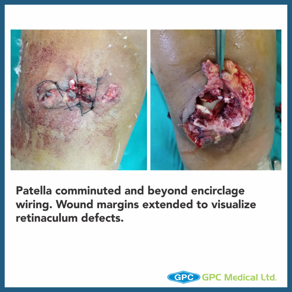
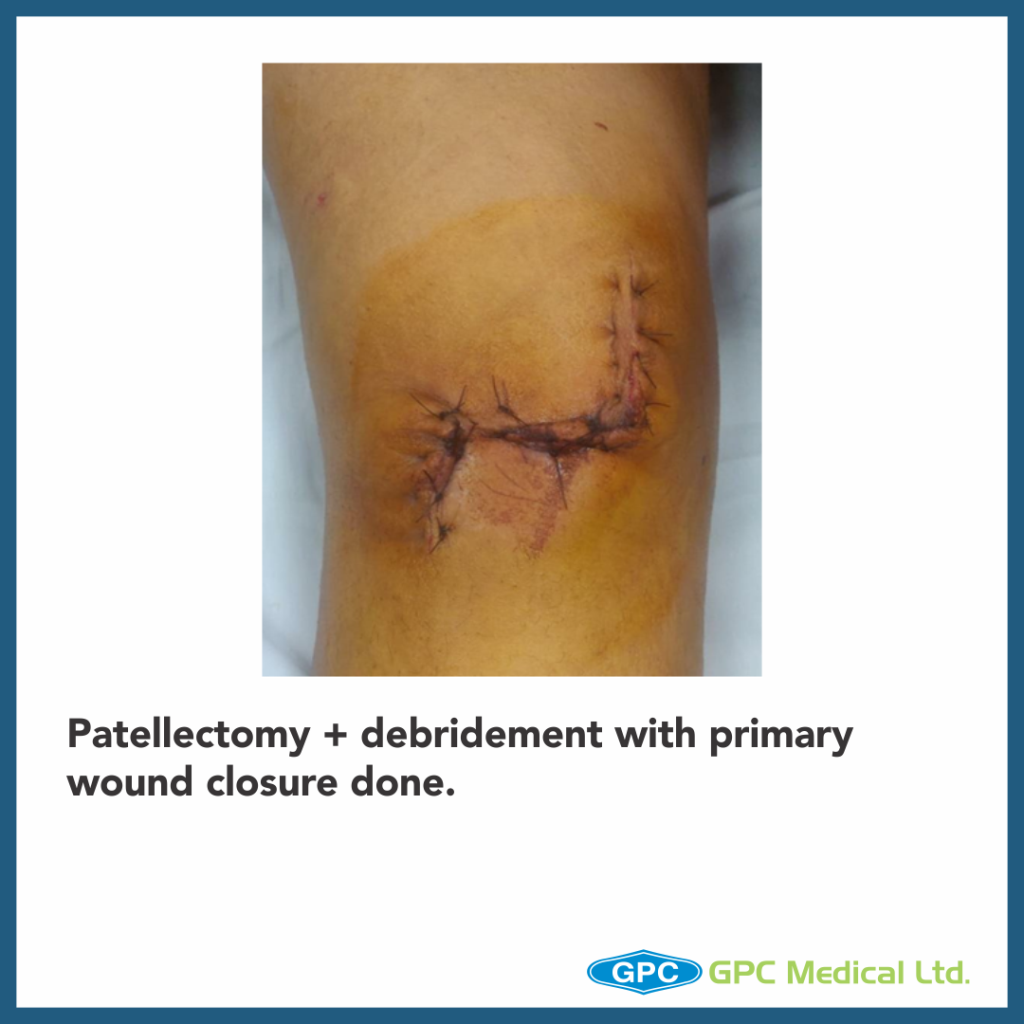
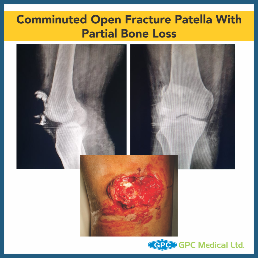
Today, we’re going to talk about Orthopaedics beyond the implant surgery. Here is one of the cases of comminuted fracture of patella, with partial bone loss. Patellectomy was performed. All that was needed was cerclage wire, of course with excellent surgical skillset.

Clinical History

  • 27 year old male
  • History of RTA
  • Presented on the day of Injury
  • Wound over right knee

Pre-operative Plan

  • Encirclage wiring + figure of eight wiring + removal of small bony fragments
  • Back up plan: total patellectomy
  • Patellectomy is still considered as the last reserve in treatment of certain conditions including comminuted fractures, advanced chondromalacia or osteoarthritis, infections, and tumoral conditions.
  • Problems associated with this procedure are:
    • Decrease in the moment arm of the extensor mechanism
    • Alteration of the forces acting on the tibiofemoral joint and instant center of motion
    • Limitation of range of motion
    • Anterior instability
    • Loss of protection of the trochlea from injury
    • Poor cosmesis
  • Extensor mechanism may be repaired in a transverse or longitudinal fashion.
  • Transverse repair only 15% of additional force was required to extend the knee after a transverse repair compared with 30% after a longitudinal repair.
  • The main objection to a transverse repair is that complete flexion of the knee is limited because of the decrease in the length of the quadriceps-patellar tendon unit.
  • An additional criticism is the longer period of immobilization that is needed to protect the tension on the suture line.(Transverse repair)

To Overcome Certain Challenges of Patellectomy

  • Some parts of the quadriceps muscle are advanced over the site of the excised patella.
  • These techniques provide relatively better cosmesis and better protection of the trochlea from injury.
  • Anterior instability and lateral subluxation of the tendon also are avoided when these techniques are used.

Miyakawa technique of patellectomy

  • In this technique, a strip of quadriceps tendon is pulled distally to fill the void that was left by the removal of the patella; the vastus medialis and lateralis then are advanced over the site of the excised patella.

Gunal technique- Patellectomy + vastus medialis obliquus advancement technique.

  • In this technique, the defect is closed longitudinally and the vastus medialis obliquus is advanced distally and laterally and distal 1 cm and is plicated to increase the angle of insertion in the sagittal plane
  • Patella is an important part of the exterior mechanism and should be preserved if possible; when patellectomy is indicated, it seems logical to combine patellectomy with reinforcement techniques.
  • For reference

Inter-Trochanteric Fractures Series – PART 1

From today, we’re starting the series of InterTrochanteric Fractures.
Today we’re talking about Significance of maintaining Medial Calcar in IT Fracture Management.

CASE 1

PRE OP X-RAY

Clinical Presentation:
– 80 years male.
– Slip and fall at home.
– Severe pain in left hip with inability to stand

Radiological investigation:
– X-Ray pelvis with both hips – Anteroposterior view

Diagnosis:
– Unstable intertrochanteric fractutre.

CASE 2

PRE OP X-RAY

Clinical Presentation:
– 84 years female.
– Fall in bathroom.
– Severe pain in right hip with inability to stand

Radiological investigation:
– X-Ray pelvis with both hips – Anteroposterior view

Diagnosis:
– Unstable intertrochanteric fractutre.

Surgical Principles and lacunae in Management

  • Standard of management for unstable intertrochanteric fracture is proximal femoral nailing because of its superior biomechanical properties.
  • Lacunae
    • Inability to obtain reduction with non maintenance of medial calcal is unacceptable.
    • It can be obtained by proper patient positioning and closed manipulation (increasing limb adduction in Case 1).
    • Carry out open reduction if result is unsatisfactory
    • Appropriate maintenance of medial calcar with centre-centre placement of spiral blade leads to proper directional controlled collapse on weight bearing and hence fracture union.
    • While poor reduction inturn leading to non centre-centre placement of spiral blade increases chances of fracture non union and blade cut-out rates.

Fracture Neck of Femur Series – PART II

This is part 2 of the Fracture neck of femur series- Understanding basics of screw fixation #NOF

We reduce and fix displaced NOF fractures under the age of 65

Except when
– Presence of significant co-morbidity or pre-existing disability
– True age or physiological age is 65 or older.
– Delayed presentation of displaced fracture (> 3 days).
– Pre-existing hip pathology

Under these exceptions, we would consider a Hemiarthroplasty or THR.

Common Issues

  • Screws biased towards anterior half of head
  • Inferior screw too far from calcar
  • Screws too short and not reaching sub-chondral bone or penetration of articular cartilage

Reduction Method

  • The most successful method of addressing this multiplane displacement is to initially restore length.
  • Applying increasing traction until the cortical break along the inferior neck of the proximal and distal segment are at the same vertical position.
  • Once length has been achieved, internal rotation will appose the fractured surfaces while correcting anterior angulation.
  • Reduction of this anterior angulation may be facilitated with manual pressure applied to the anterior femoral neck, combined with a lateral distractive force applied to the femoral shaft.
  • Once the sagittal plane angulation has been corrected, adduction of the leg will generate tension within the abductor musculature, which will lock the reduction in place.

Three-screw pattern, each screw has specific mechanical function

  • Inferior screw- along the inferior cortex in the anteroposterior projection and centrally on the lateral projection. This position will optimally resist inferior displacement and varus collapse while the femoral head is loaded in a standing position.
  • Posterior screw is placed parallel to the first along the posterior cortex and centrally on the anteroposterior image. This pin will resist posterior displacement and anterior angulation while the patient is rising from a seated position.
  • The final screw should be placed anteriorly on the lateral view and centrally on the anteroposterior view.
  • Traction is released, and gentle impaction along the axis of the pins is performed manually.
  • Further screw tightening may be required to minimize postoperative collapse and resultant symptomatic hardware.
  • Thread length should be chosen so that threads do not cross the fracture, allowing for postoperative impaction.
  • Continuous fluoroscopy during internal and external rotation, in both anteroposterior and lateral projections, may aid in this assessment of containment of all of the screws within the femoral head

Pearls

  • Strive for cortical support of implants in osteopenic bone, using the inferior and posterior cortices of the femoral neck.
  • When the starting point of a guidewire is correct but angular change is necessary, the pilot hole may be expanded with a cannulated drill bit, which will allow angulation of the guidewire and improved positioning.
  • When open reduction is required, a drill hole placed in the anterior neck serves as an excellent seating point for a pointed reduction clamp.
  • While performing antegrade or retrograde nailing in the setting of an associated femoral neck fracture, generous overreaming of 1.5 to 2 mm will reduce the force necessary for nail insertion and, in turn, reduce the risk of femoral neck displacement

GPC Screws

  • 7.3 mm Screws
  • 2.8 mm Guide wires
  • 16 mm/32 mm thread
  • Titanium & Stainless steel (S.S. screws avoided due to need of MRI in future)
  • Self-drilling & self-tapping

Fracture Neck of Femur Series – Part 1

Fracture Neck of Femur Series

Part 1 – Understanding Case Failure

PRE OP X-RAYS

Femur Fracture

Clinical Presentation

  • 64 year old female
  • Osteoporotic
  • Fall at home while having bath
  • Severe hip pain and inability to stand

Radiological investigation

  • Anteroposterior View
  • Lateral View

Diagnosis: Fracture neck of Femur

Intraoperative reduction unsatisfactory
Screw fixation of malreduced fracture
Two week postop xray
6 week post op X-rayTrochanteric fracture, No trauma-fixation failure

Surgery Principle and Lacunae in Management

  • Femoral head salvage surgery with reduction of fracture and internal fixation with CC screw
  • Lacunae
    • Residual varus is absolutely not acceptable.
    • Failure of closed reduction should have prompted for open reduction or change of surgery plan to hemiarthroplasty.
    • Poor reduction left a very small corridor to pass the three screws, therefore all three screws were cramped together.
    • The varus reduction and the lack of spread of the screws add up to make this a very unstable construct.
    • Fracture found to be a propagation of the three screw holes, two of them were very close and joined up as one big oblong hole.

Distal Humerus Fracture – Case Discussion

In this video you will see a very interesting trauma case of distal humerus fracture. A special mention to our surgeon from Malaysia, who was kind enough to share the details with us.

A 24 year old female had a road traffic accident with injury to the left elbow. It was a closed fracture without any neurovascular deficit.

Case Discussion

  • 24 year old female
  • Thinly built
  • Road traffic accident with injury to the right elbow
  • Closed fracture without any neurovascular deficit

Radiological investigation

  • Anteroposterior view
  • Lateral view
  • CT scan
  • Traction View X-ray (optional)

Fixation Principle

  • Stable internal fixation of the articular surface
  • Restoration of articular axial alignment
  • Stable internal fixation of the articular segment to the metaphysis and diaphysis
  • Early range of motion of the elbow


Rationale

5.0mm cannulated screw in fixation of medial column

  • Good purchase, better compression, and stable fixation
  • Only when medial column is not comminuted
  • Less dissection, shorter operative timing
  • Less implant impingement
  • Avoid double-plate stress-riser effect
  • Much cheaper than plate and screws


Patient developed olecranon bursitis with screw prominence at 6 months


Use of screw for fixation of olecranon osteotomy

  • Large-diameter screw threads may engage ulnar diaphysis prior to full seating of screw head
  • “Bite” of screw may be strong without full compression.
  • Beware of the bow of the proximal ulna, which may cause a malreduction of the tip of the olecranon if a long screw is used.
  • Eccentric placement of screw may be helpful
  • Careful scrutiny of lateral radiograph important to assure full seating of screw head
  • Long screw may be beneficial for adequate fixation
  • Short screw may loosen or toggle with contraction of triceps against olecranon segment


26 months


Case Discussion – A 7 year old male child presented to the ER with pain over the right thigh along with swelling & inability to bear weight.

Case Discussion

A 7 year old male child (weight 23 kg)

  • History of fall from swing
  • Presented on the day of injury
  • Pain over right thigh with swelling and inability to bear weight

Pre-Op X-rays

Q. What would be the ideal treatment plan?

  • Closed reduction and Hip Spica
  • TENS +/- single leg spica/POP Splintage
  • TENS/ Ender’s nailing(Stack)
  • Open reduction and plating
  • Closed Reduction + MIPPO

A. Even though the treatment options can be debatable. However, in this case the patient was treated with Closed reduction+ MIPPO.

Fracture geometry shows the fracture to be unstable type. With conservative treatment patient will be immobilized in hip spica for long duration with high possibility of limb shortening.

With TENS nailing similar problems may be seen however probability of such complications will be less than conservative treatment.

Closed reduction and MIPPO is a stable fixation method with maximizing biologic fracture healing potential. The fracture anatomy is left undisturbed and plate fixation is done in bridging mode with plate acting as an internal fixator/extramedullary splint akin to external fixator. In such cases the intermediate fracture zone is left untouched. Anatomical reduction of intermediate fracture fragments is not necessary. Slight overlap is acceptable with remaining growth potential of the child. Overzealous traction to maintain limb length might result in traction injuries to neurological tissues.

In comparison to TENS it provides a stable fixation method with less chances of rotational deformity.

Open plating of pediatric femoral fractures have been associated with femoral overgrowth as a result of periosteal stripping.


Case Discussion – Elderly male patient visited the ER with pain over the left hip with swelling & bruising. He had a fall at home, the same day.

Check out the pre-op & post-op X-rays.

Join us for discussion & find out the answers to the following questions:

Q. What are the issues with this fixation?

Q. Which factors lead to these issues?

Q. How to troubleshoot these factors & achieve ideal implant positioning?

Q. What can be the post-operative management protocol?

Hip Fracture - Orthopedic Surgery - Internal Fixation

Q. What are the issues with this fixation?

Fracture reduction is inadequate, fracture is fixed in varus. High probability of implant failure in such cases. Over time such fractures go into varus collapse and either there is a blade cut out from the femoral head or implant failure and breakage.

Q. Which factors led to these issues during fixation?

Entry point was lateralised and the guide wire passed through the fracture site. Eventually the nail was passed through the fracture site and the implant pushed the femoral head into varus position.

Q. How to troubleshoot these factors to achieve ideal implant positioning?

Sometimes the nail pushes the femoral head into varus position, especially when the nail goes through the fracture gap at the trochanteric region with inadequate reaming at the entry site. Some surgeons always ream the entry portal by hand without using the power tool. With proper or slight over-reaming, varus tilt of the head and neck fragment can be prevented.

In some cases guide pin abuts against the iliac crest and it goes towards varus position. It is more common in patients with short height. It is recommended that the torso of the patient should be pushed towards the opposite side.

Q. Any changes in post-operative management protocol in such cases?

Weight bearing is delayed till there are signs of callus formation. Number of hospital visits increase and monthly/bimonthly check x-ray are required till significant consolidation of fracture is seen. These constructs are biomechanically unstable so weight bearing is to be done in gradual manner. Family and patient needs to be counselled about chances of implant failure and increased pain at the affected site requiring surgical intervention.

A case study on Distal Radius Fracture Surgery and Post-operative Complications

Q. A 52-year-old man, presented to emergency department with pain over right wrist joint following road traffic accident. X-ray revealed a comminuted distal radius fracture. Patient underwent open reduction internal fixation. Immediate postoperative radiographs are shown here. What complication is most likely to occur in this patient in the early postoperative period?

A. The first concern is that there are chances of screw penetration into the wrist joint due to distalization of the radius plate up to the volar rim. The implant used in this case is a variable angle plate, a screw can be directed away from the fracture line in this case while using this implant. The longer screw should have been used to fix the radial styloid fragment. The radial styloid fragment has two screws but both of them are close to the fracture line. Wrist joint violation by screw penetration will severely hinder the joint range of motion due to pain and cause early joint degeneration.

Distal Radius Fracture Postoperative Radiographs

If we consider the screw to be adequately placed, A well-fixed distal radius fracture using locking plates can be mobilized in the early postoperative period and don’t require splinting support thus giving an early range of motion and early functionality to the limb. The dorsal fragment was not fixed in this patient, so to achieve stability there is need of splintage for 2-3 weeks for the fracture to consolidate before starting the mobilization process. Such fracture requires dual plating for fracture fixation. Longer volar plate and a shorter dorsal plate would have been ideal for such a scenario. Other option would have been to reduce the dorsal fracture by indirect reduction methods and temporarily fixed with K wire and to be fixed with screws after volar plating is completed. In this second case, splintage would have been required but the fracture reduction will be more satisfactory than the present scenario.

For GPC Medical, the priority is your health and quality of your life

Orthopedic implants are the devices that are surgically inserted into the body and are intended to reinstate function by restoring or strengthening a damaged structure. Global Orthopedic Implants Market is booming internationally. The orthopedics market covers a wide range of medical devices, from bone cement and surgical power tools to joint replacement implants and prosthetic limbs, all of which aim to provide patients with increased mobility. Driven largely by the aging population and patients’ desire to maintain an active lifestyle, the global orthopedics market was valued at $52.7B in 2017. GlobalData expects this market will grow to $66.3B in 2023 at a steady Compound Annual Growth Rate (CAGR) of 3.9%.

GPC Orthopedic Implants & Instruments are globally reputed for their high quality, precision and durability. Our Orthopedic Surgery Instruments & Implants, in large quantities are regularly exported to many different countries including those in Europe. Meeting customized requirements of Orthopedic Surgery Instruments and Implants is our specialty. Most of our Orthopaedic Implants including spinal system, intramedullary interlocking nail, locking/non-locking bone plates & screws are US FDA 510(K) approved. GPC is the only Indian Company so far to have achieved this distinction in the field of Orthopaedic/Medical Equipment.

Our wide range of Orthopaedic Implants & Surgical Equipment includes Bone Screws, Plates, Intramedullary Nails, Maxillofacial Implants, Knee Implants, Hip Prosthesis, Spinal Implant System, Locking Plate System, External Fixators, Power Tools, General Orthopedic Instruments, Instruments Sets, Surgical Drills, Arthroscopy Instrumentation, etc. etc.

Orthopedic Implants - GPC Medical Ltd.


Scope of orthopedic Implants

GPC Medical Orthopedic Implants occupy a lion’s share in the international orthopedic implant market. Orthopedic implants are associated with the musculoskeletal system. For instance, to treat a backache orthopedic implants such as bone plates or bone screws are utilized in spinal fusion surgery. Fixation of damaged bone fragments as well as implant components is used for hip, knee and any other joint replacement. The material applied in orthopedic implants must be biocompatible to avert rejection by the body.

Implant Potential

Orthopedic implants are manufactured to replace or support a missing or damaged bone or joint. These implants are intended to treat the deformities, stabilize body posture, and restore the normal skeletal function. The orthopedic implants market has witnessed a shift from conventional surgical procedures to the use of modern fixation and prosthetic devices. The demand for orthopedic implants has increased significantly, owing to rise in geriatric population that increases the risk of osteoporosis, osteoarthritis, technological innovations in the orthopedic implants, and other musculoskeletal disorders. GPC Medical knows how to keep up with technological demands and adapt via their innovation laboratories with experienced surgeons and engineers.

Orthopedic Implants Market Synopsis

Almost every orthopedic implant present in the market in the current scenario, come under the class III category, which is the most scientifically meticulous classification of medical devices. Individuals facing immobility issues due to terrible accidental or traumatizing cases have no other alternative but to opt for major joint replacement surgeries. This can eventually provoke a radical change in people’s quality of life by relieving pain and reviving their physical independence.

Key Segments

The global orthopedic implants market is segmented based on product, biomaterial, and geography. Based on product, it is classified as reconstructive joint replacements, spinal implants, dental implants, trauma & craniomaxillofacial, orthobiologics, and others. The reconstructive joint replacements market is further divided into the knee, hip, and extremities. Spinal implants are further categorized into spinal fusion devices, non-fusion devices/motion preservation devices, and invasive spinal bone stimulators. Dental implants are further bifurcated into root form dental implants and plate form dental implants. Trauma & Craniomaxillofacial are further categorized into screws, plates, staples, and others. Orthobiologics are further classified into allografts, bone graft substitutes, and cell-based matrices. Based on biomaterials, it is categorized into metallic, ceramic, polymeric, and natural biomaterials. Among biomaterials, the metallic segment occupied dominant share in 2016 and is expected to maintain this trend during the forecast period, owing to their exclusive load-bearing properties.

Factors Leveraging Market Share

Rising geriatric population susceptible to increased risk of osteoarthritis, osteoporosis, and obesity lowered bone density; increased body mass index and other musculoskeletal disorders are the vital factors driving the growth of the global orthopedic implants market. Furthermore, a significant rise in the number of sports-related injuries and road accidental cases are another contributors stimulating market growth. Besides, technological advancement, innovative procedures, and swiftly growing healthcare industry further incite the growth of the market.

Moreover, the launch of internal fixation devices and biodegradable implants holds the higher potential for future prospects of orthopedic implants. However, unfavorable reimbursement policies, rigid guidelines authorization procedures, the high cost of the treatment might hamper the market development.

Additionally, the risk associated with orthopedic implants including implants coming loose or breakage in the bone causing painful inflammation and infections together with the possibility of implant rejection could also serve major roadblock to market expansion.

Technological approach GPC Medical

The impact of the driving factors is expected to surpass the effect of restraints. Moreover, GPC Medical’s increase in R&D in orthopedic implants and the untapped emerging economies are expected to provide new innovative products to orthopedic implants market in the near future.

Future of Global Orthopedic Market

Moving into 2018, there are several areas to watch that GlobalData anticipates will affect the orthopedics markets. Patient-specific implants are expected to drive growth in the hip and knee markets, as is patient-specific instrumentation (PSI), a tool developed to improve accuracy in total knee arthroplasty procedures by customizing the surgical cutting blocks to the patient’s anatomy. Similarly in the vein of personalized healthcare, 3D printing in a variety of orthopedic markets, including spinal intrabodies, craniomaxillofacial implants, and prosthetic devices, will advance physicians’ ability to provide customized solutions to patients. GPC Medical Limited aims to absorb all these changes to make availability of the best quality implants based on these technologies.

A Futuristic Approach

GPC Medical believes in growth and advancement. This is why it keeps a futuristic approach with orthopedic implants. Minimally invasive and robotic surgical systems are a growing trend in orthopedic surgeries, and major orthopedic players, including Medtronic, Stryker, Smith & Nephew, and DePuy, are taking steps to ensure they remain competitive with their Mazor X, Mako, NAVIO, and PUREVUE systems, respectively. Lastly, a shift in the US towards value-based healthcare, including Medicare’s Comprehensive Care for Joint Replacement (CJR) model, will continue to impact both orthopedic manufacturers and physicians, as will the move towards outpatient procedures in ambulatory surgical centers (ASCs). GPC Medical Limited is not ready to stop at any point, as we keep adopting these technological advancements.

Orthopedic Implants Manufacturing Company

Why GPC Medical Limited?

GPC Medical Limited is the oldest orthopedic implants manufacturer and international supplier. Our technology labs remain busy in innovating and manufacturing Trauma Implants with modern technology machines viz. Sliding Head Turning Center, Multiple Axis VMC, CNC Wire Cutting, CNC Deep Hole Drilling, Mechanized sharpening & surface finishing machines.

Apart from our wonderful market acceptance along with the biggest manufacturing portfolio of over a thousand products in growing a worldwide market, we have built relationships based on trust and reliability. We follow internationally standardized protocols with precision throughout the manufacturing and innovating process.

GPC Medical Futuristic Facilities

GPC Medical Limited Manufactures Orthopedic Implants and Instruments product ranges which are packed in packages manufacture from the extraordinary material which is best and suited for orthopedic implants manufacturing process. Further, implants and instruments are marked with Cat Numbers, Batch Numbers and complete description with computer laser marking techniques. GPC medical remains conscious about its internationally renowned reputation and stringent compliance of international standards.

US FDA 510(k) Approved Orthopedic Implants - GPC Medical Ltd.

GPC Medical Ltd. – US FDA 510(k) Approved Orthopedic Implants