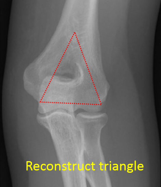
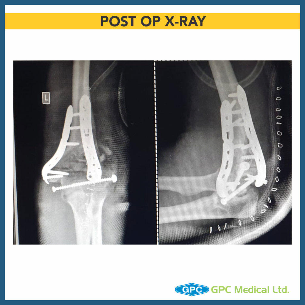
Elbow Reconstruction – Understanding Failure (PART 2)

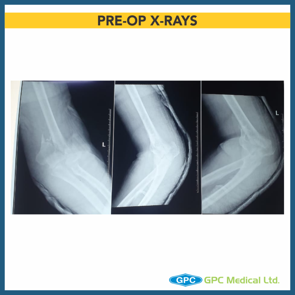
Continuing on last post on elbow reconstruction, today we’re going to talk about the contingency plan i.e. when the surgeon attempted to reconstruct column, but failed to achieve intra-articular fragment reduction.

Clinical Presentation

  • 29 year old female
  • Thinly built
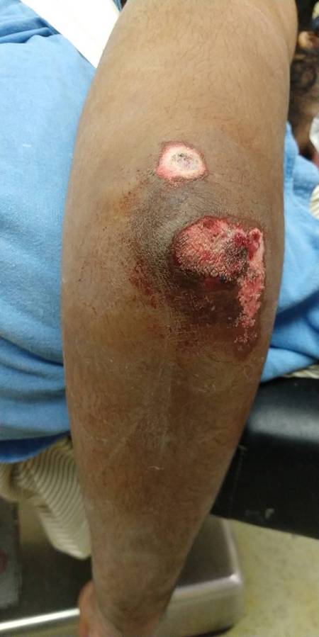
  • Road traffic accident with injury to the left elbow
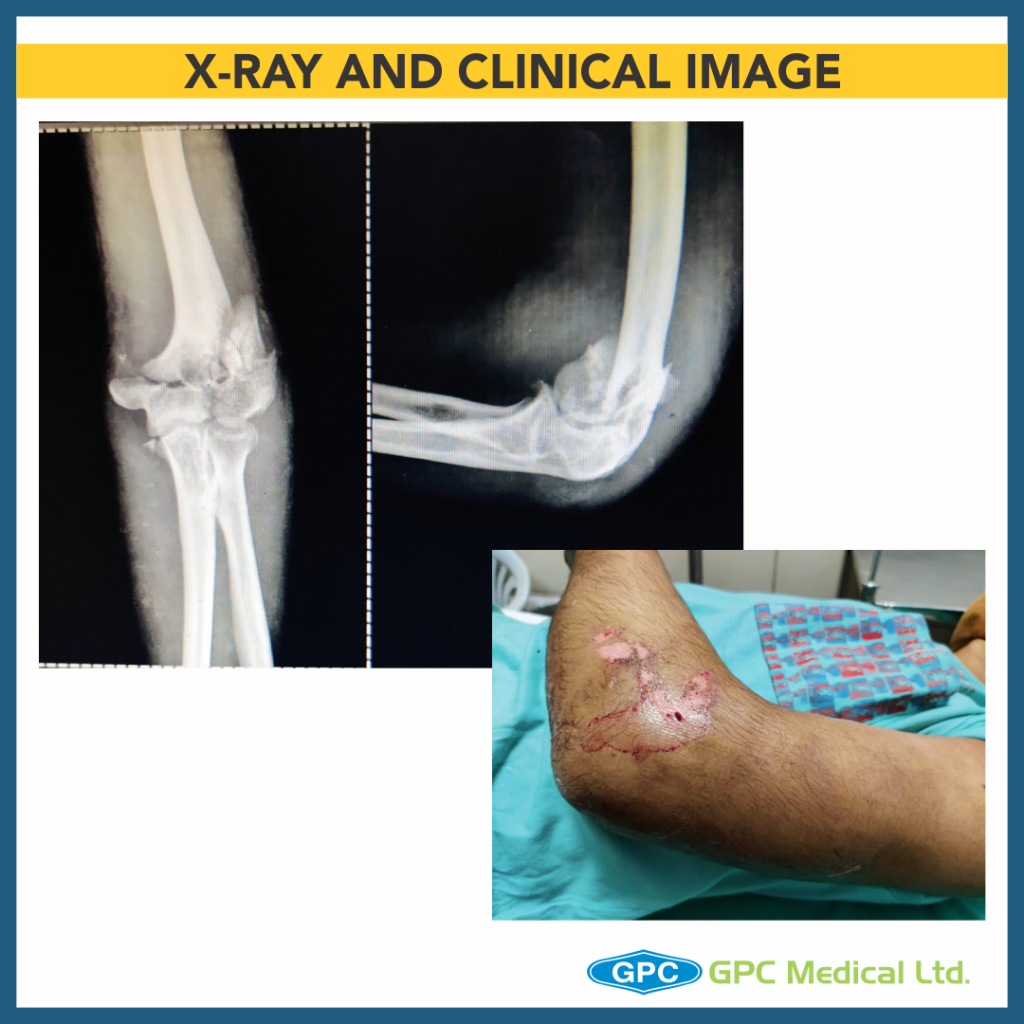
  • Trauma to the skin (abrasion) overlying the fracture
  • Closed fracture without any neurovascular deficit
  • Co-morbidities – None

Investigations

  • X-ray of the suspected region- pelvis/chest/spine screening/limbs
  • X-ray of affected region- anteroposterior/ lateral/ traction views (author preference)
  • CT scan of elbow (cost constraint)- gold standard
  • Blood workup for surgical fitness

What should have been done?

  • Author’s preferred method- discussed in one of earlier discussion on distal humerus fractures.
  • Simple plan
    • Olecranon osteotomy
    • Intercondylar partially threaded cancellous screw (lag effect)
    • Temporary column fixation
    • Medial column single screw fixation
    • Lateral column fixation by 3.5 mm LCP/Lateral Distal humerus plate
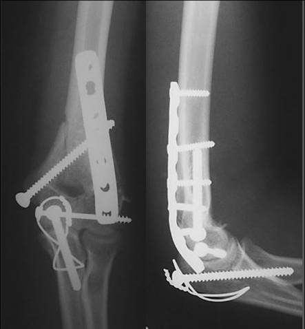
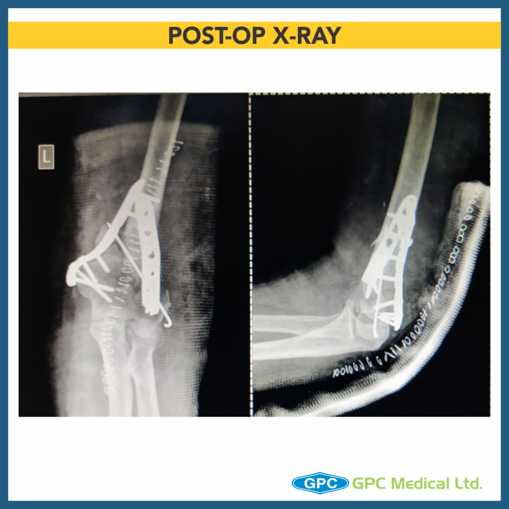
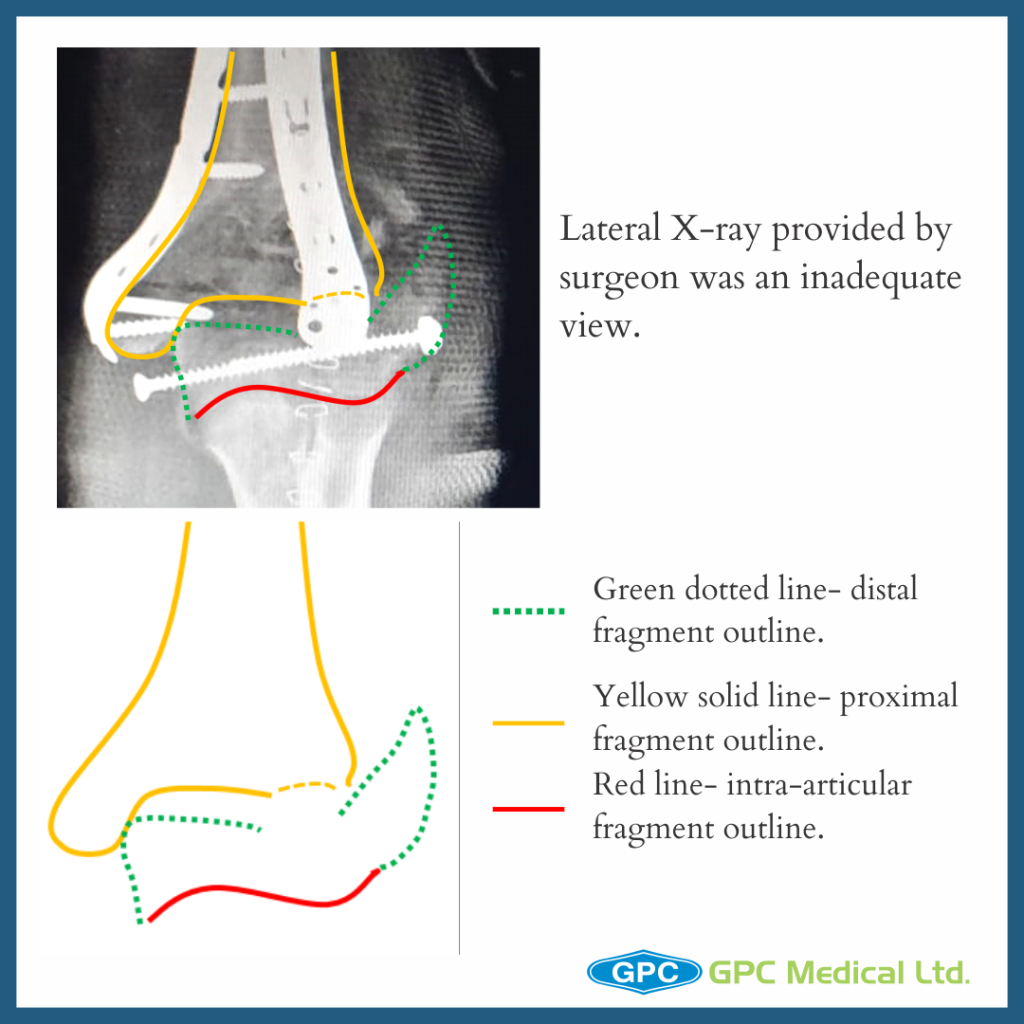
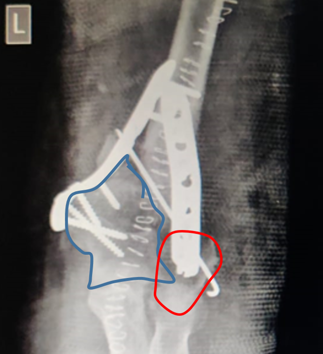
Fixation Issues

  • No compression in between two condylar articular fragments
  • Medial column not restored
  • Anterior angulation of distal humerus not maintained
  • Lateral distal fragment- capitullum is fixed in rotation
  • Short screw length
  • Slab application (length) is not appropriate for such injuries
Why does one land in such issues?

  • Pre-op formulate a plan to fix such complex trauma case
  • Follow the plan, ensure each step is correctly addressed
  • Back-up plan should be there in case difficulty/ problem in original plan (author would have to drop idea of single medial screw and go for medial plate due to comminution)
  • Critical analysis of the postop case to ensure adequate results in next case
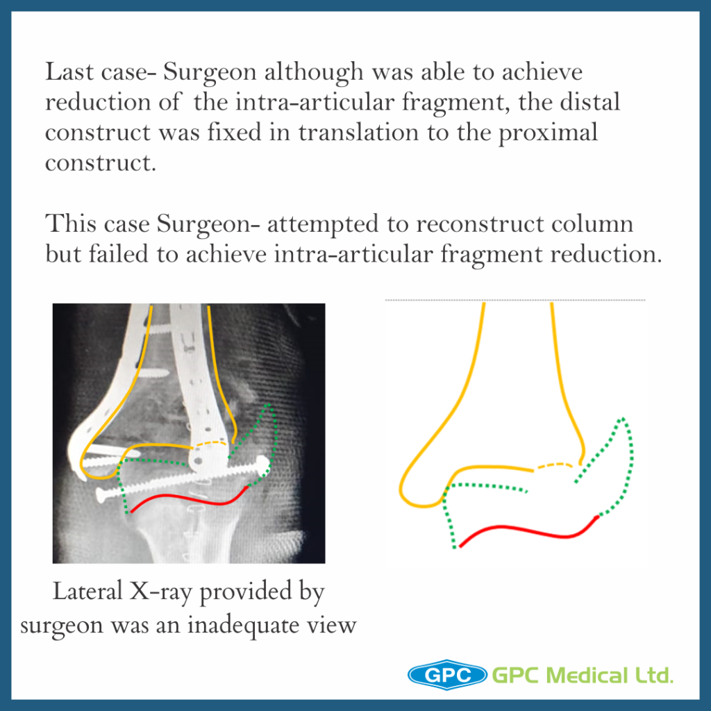
Last Case Discussion

  • Pre-op & contingency plan
  • Importance of exposure and different methods
  • C-arm visualization
  • Patient expectancy from injury
  • Strongest bone lies along medial and lateral columns and therefore the implant should be placed here
  • Intra-articular anatomical reduction and fixation by lag screw placed from medial to lateral direction
  • With normal 40° anterior angulation of the condyles and humeral shaft restored, lateral plate is positioned posteriorly & medial plate in saggital plane along the medial border.
  • Screws in the distal fragment need to be as long as possible,engaging as many fracture fragments as possible, ensuring screw tip is not impinging into the joint
  • Medial and lateral column screws should “interdigitate“ such that they have hold on opposite column fracture fragment 
  • Check ROM and fracture fixation stability on the OT table before closure

Elbow Reconstruction in Trauma Cases – PART 1

Today, we’re going to talk about Elbow Reconstruction in trauma cases. The main principle which we have to keep in mind when reconstructing the elbow is- stable internal fixation of articular surface & also stable internal fixation of the articular segment to the metaphysis & diaphyseal fragments.

Clinical Presentation

  • 34 year old male
  • History of RTA
  • Presented on the day of Injury
  • Pain and swelling over left elbow with abrasion and bleeding from the wound
  • Difficulty in elbow movement
  • Co-morbidities – None

Investigations

  • X-ray of the suspected region- pelvis/chest/spine screening/limbs.
  • X-ray of affected region- anteroposterior/lateral/traction views (author preference)
  • CT scan of elbow (cost constraint)- gold standard
  • Blood workup for surgical fitness
  • Additional test as required for co-morbidities if any

Clinical Evaluation

  • Soft tissue envelope
  • Neurological status distal to injury
  • Vascular status distal to injury

Goal and Principle of Surgical Fixation

Goal of treatment

To achieve stable fixation, union of fracture fragments with restoration of function of elbow.

Principles of fixation

  • Anatomical reduction of intra-articular fragments
  • Preservation of blood supply
  • Stable fixation
  • Early and safe mobilization

Importance of Surgical Plan

  • Surgeon needs to pre-formulate a surgical fixation plan and the necessary approach for adequate exposure.
  • In addition to the above planning, a backup/contingency plan also needs to be formulated in such complex trauma cases.

Surgical Issues

  • Principle of stable fixation constitute
    • Stable internal fixation of articular surface
    • Stable internal fixation of the articular segment to the metaphysis and diaphyseal fragment
  • The surgeon although was able to achieve reduction of the intra-articular fragment, the distal construct was fixed in translation to the proximal construct.
  • Different exposures gives good to complete visualization of articular surface
  • Choose the exposure depending upon
    • Surgical experience
    • Type of implant
    • Patient factors- skin condition/age/fracture pattern
    • Future need of elbow arthroplasty
  • Author’s preference
  • Olecranon osteotomy (fixation with TBW/ Long screw+ SS wire)
  • Byran-Morrey Approach (Triceps-sparing postero-medial approach)
    • Midline incision
    • Ulnar nerve identified, mobilized and latex loop placed
    • Medial edge of triceps and distal forearm fascia elevated as single unit off olecranon and reflected laterally
    • Resection of extra-articular tip of olecranon(better visualization, later helps during ROM exercises)

Adequate Visualization on C-arm During Surgery

At 90° flexion: The olecranon overlaps only a small part of the medial joint surface

  • If elbow flexion is increased to 110°- free and complete joint surface is seen on lateral radiograph
  • It is therefore important to have proper patient positioning allowing elbow movement and facilitate radiography.

Pre-op Counselling

What patients may expect?

  • Loss of 15-25° of flexion and extension
  • Maintain most of supination and pronation
  • Decrease in muscle strength (triceps)
  • Implant impingement (especially olecranon fixation implant)
  • Complications associated with the procedure

Fracture Olecranon Management with TBW (Tension Band Wiring)

Check out our latest blog post where we are putting a spotlight on tension band wiring for Olecranon Fractures with relative case studies and imagery. We also discuss concerns relating to Olecranon Fracture and K-wires and discuss possible alternatives for the same.

Clinical History

  • 17 year old male with history of RTA
  • Pain, swelling with restricted movement of left elbow
  • No distal neurovascular deficit

Treatment Planning

  • Xray- Comminuted fracture olecranon
  • Ideal surgical treatment option- Olecranon plating
  • Cost concerns- TBW
  • The principles of tension band wiring is conversion of tensile forces into compression forces.
  • In the olecranon, the figure-of-eight wire loop lies on the posterior surface acts as a tension band during elbow flexion.
  • For success of tension band wiring in olecranon fracture, the anterior cortex cannot be comminuted and must provide a buttress to allow compression.
  • Multifragmentary olecranon fractures cannot be fixed with tension band wiring.

Issues with fixation

  • Fracture reduction is unsatisfactory- inadequate hold of K wire on one fragment leading to displacement.
  • No transcortical fixation of K wires
  • Triceps tendon shadow visible under the Wire loop. Wire loop should pass under the triceps to provide compression of fracture during loop tightening.
  • K wires not sitting on the proximal fragment.
  • Wire loop placement on distal fragment should pass through centre of ulnar distal fragment.
  • Comminuted olecranon fracture [Types IIB, Mayo Classification] a posterior splint with the elbow in a semi-flexed position applied for a period of 2–3 weeks to prevent fracture collapse and displacement. In this case its slab in 90 degree flexion.

Olecranon fracture surgical management concerns

  • High chance of needing a secondary procedure for hardware removal due to irritation.
  • Frequently a loss of terminal elbow extension of 10-15 degrees without any functional deficit
  • Fractures of the olecranon are intra-articular injuries and require anatomic reduction.  Be careful to reduce the bare area of the articular surface.
  • Do not malreduce the fracture and narrow the distance between the coronoid process and tip of the olecranon.

Sullivan CW, Herron T, Hayat Z. Olecranon Fracture. [Updated 2020 Nov 1]. In: StatPearls [Internet]. Treasure Island (FL): StatPearls Publishing; 2020 Jan-. Available from: https://www.ncbi.nlm.nih.gov/books/NBK537295/

Distal Humerus Fracture – Case Discussion

In this video you will see a very interesting trauma case of distal humerus fracture. A special mention to our surgeon from Malaysia, who was kind enough to share the details with us.

A 24 year old female had a road traffic accident with injury to the left elbow. It was a closed fracture without any neurovascular deficit.

Case Discussion

  • 24 year old female
  • Thinly built
  • Road traffic accident with injury to the right elbow
  • Closed fracture without any neurovascular deficit

Radiological investigation

  • Anteroposterior view
  • Lateral view
  • CT scan
  • Traction View X-ray (optional)

Fixation Principle

  • Stable internal fixation of the articular surface
  • Restoration of articular axial alignment
  • Stable internal fixation of the articular segment to the metaphysis and diaphysis
  • Early range of motion of the elbow


Rationale

5.0mm cannulated screw in fixation of medial column

  • Good purchase, better compression, and stable fixation
  • Only when medial column is not comminuted
  • Less dissection, shorter operative timing
  • Less implant impingement
  • Avoid double-plate stress-riser effect
  • Much cheaper than plate and screws


Patient developed olecranon bursitis with screw prominence at 6 months


Use of screw for fixation of olecranon osteotomy

  • Large-diameter screw threads may engage ulnar diaphysis prior to full seating of screw head
  • “Bite” of screw may be strong without full compression.
  • Beware of the bow of the proximal ulna, which may cause a malreduction of the tip of the olecranon if a long screw is used.
  • Eccentric placement of screw may be helpful
  • Careful scrutiny of lateral radiograph important to assure full seating of screw head
  • Long screw may be beneficial for adequate fixation
  • Short screw may loosen or toggle with contraction of triceps against olecranon segment


26 months


What is the treatment plan? Whether to operate or manage the patient conservatively?

A five year old child presented with tenderness over the lateral aspect of the elbow, with bruised skin & swelling. The radiograph shows fracture of the elbow with displaced lateral condyle.

Q. What is the treatment plan? Whether to operate or manage the patient conservatively?

Elbow Fracture Radiograph

A. Since it is a type 4 displaced fracture, the patient was treated by open reduction & internal fixation with K-wires. Here is the post op X-ray.