Distal Tibia & Fibula Fracture

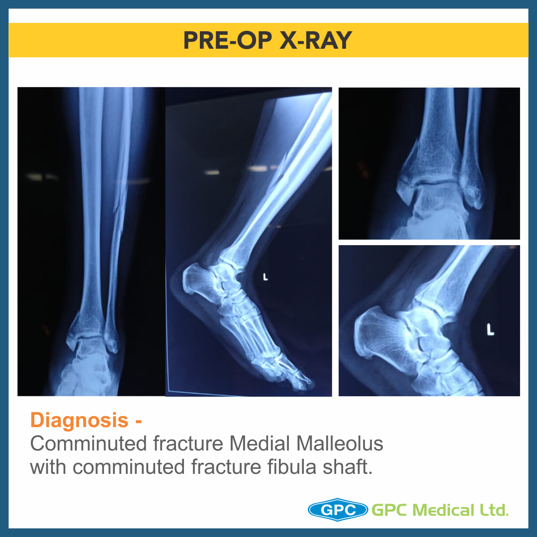
Today, we are going to discuss a very interesting case, which one of our surgeons came across last week. A 56 year old male presented with swelling around ankle on the day of injury. He had twisted his ankle. After the X-ray, it was diagnosed as Comminuted fracture medial malleolus with comminuted fracture fibula shaft. The surgeon planned to treat him with Open reduction & internal fixation through GPC fixLOCK Medial distal tibia plate with tab & Medial malleolus screw fixation. However, intra-operatively, due to unstable ankle injury, the treatment plan was changed.

Clinical History

  • 56 year old male
  • History of twisting injury around ankle
  • Presented on the day of injury
  • Pain and swelling over left leg
  • Co-morbidities – hypertension

Diagnosis & Treatment Planning

Diagnosis

  • Comminuted fracture medial malleolus with comminuted fracture fibula shaft

Plan

  • Medial tibial plate with tab
  • Medial malleolus screw fixation + neutralization plate

Change in Treatment Planning

Diagnosis

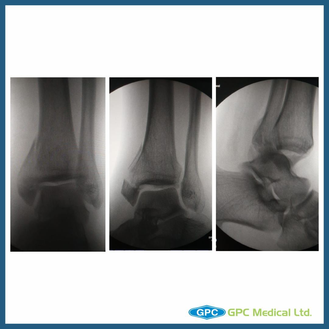
  • Comminuted fracture Medial Malleolus (Vertical and Horizontal split) with posterior malleolus fracture with Posterior Subluxation of ankle with comminuted fracture fibula shaft

Plan:

  • Medial malleolus screw fixation + neutralization plate
  • Stabilization of ankle joint

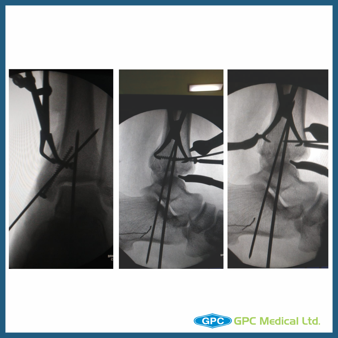
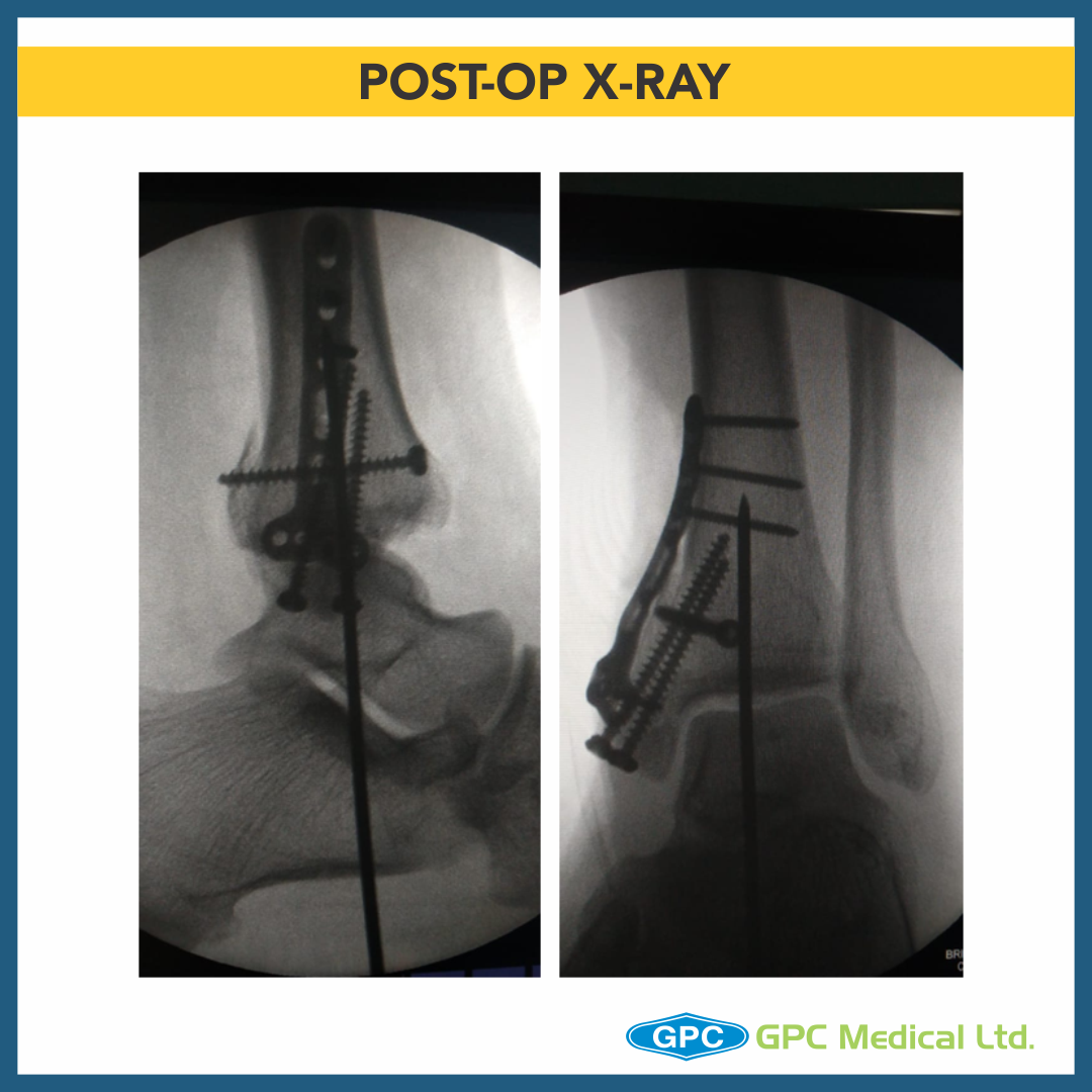
Anatomical reconstruction of joint

  • Ankle joint reduced and fixed with K wire
  • Posterior malleolus temporarily held with clamp and fixed with screw
  • Medial malleolus horizontal split fragment fixed with screws
  • Vertical split fragment had numerous small fragments
  • GPC Medical Ltd. Distal Radius T-plate used
  • Horizontal Limb of T-plate is curved to match the distal tibia and buttress the anterior and posterior aspects
  • Volar tilt of T-plate is reversed.

Minimal Soft tissue stripping
Preserved Bone Blood supply
Early return to function

Comminuted Proximal Humeral Head Reconstruction

One of our surgeons have shared a very interesting case with us. A 58 year old male presented to the ER after RTA, with pain & swelling over the left shoulder.

CASE DISCUSSION

  • 58 year old male
  • History of RTA
  • Presented on the day of Injury
  • Pain and swelling over left shoulder
  • Co-morbities – Diabetes, Hypertension

Importance of medial calcar support and long axis alignment of the fracture

  • Various studies have postulated and shown importance of medial calcar support and prevention of varus malalignment in significantly reducing the risk of fixation failure in proximal humeral fracture fixed with locking plates.
  • Inadequate fracture reduction with lack of medial bony continuity in addition to lack of medial calcar screws makes the construct as unstable with high possibility of failure leading to Nonunion, fracture redisplacement, malunion, and or implant failure & breakage.
  • With medial calcar continuity and importance of calcar screws highlighted. We would also highlight importance of primary cortical strut grafting (Allograft/autograft) during fracture fixation to give structural strength to the construct and prevent fracture redisplacement.
  • Before commencing on bone grafting, surgeon needs to reduce the comminuted facture with help of non absorbable sutures.
  • We are highlighting certain pearls from the discussion of this technique published by Panayiotis Dimakopoulos et al.
  • With this technique author has avoided implants for fracture fixation.
  • Concept of suture fixation can also be applied along with other methods discussed here in teaching Tuesday session.
  • This article reading is must to understand various fracture geometry and understanding restoration of fracture fragments.
  • The principles of suture fixation can be reapplied when using other fixation methods.
  • Invariably, the humeral head is facing superiorly with the tuberosities displaced to either side of it.
  • Two heavy nonabsorbable sutures are passed through the bone of the head fragment, 1 cm proximal to the fracture line at both the medial and the lateral border of the articular surface.
  • Additional sutures are then passed through each tuberosity fragment (or near the site of tendon insertion into the fragment in osteoporotic bone or when intensive comminution is present), and the rotator cuff tendons are mobilized.
  • Finally, two additional pairs of sutures are inserted laterally and medially through 2.7-mm drill holes in the diaphysis.
  • These sutures are then passed through the opposite tuberosity, near the musculotendinous junction, and on to the neighboring area of the articular segment (i.e., from the medial diaphysis toward the greater tuberosity and from the lateral diaphysis toward the lesser tuberosity as well as to the adjacent articular fragment).

Key points for management of comminuted fractures

  • Place sutures into the rotator cuff for control and fixation of the tuberosities.
  • K-wires were used by several authors to joystick the fragments and achieve graft position.
  • Matassi et al describes the placement of a single screw through the locking plate to push the fibula medially until it apposes the medial cortex of the humerus (to indirectly reduce the medial column) before subsequently placing several shaft screws through it.

Matassi F, Angeloni R, Carulli C, et al. Locking plate and fibular allograft augmentation in unstable fractures of proximal humerus. Injury. 2012;43:1939–1942.

Take home message

  • Appropriate fracture reduction with continued medial bony support.
  • Two calcar screws placed to support bony fragment.
  • Use sutures in bony fragments/or rotator cuff to help in reduction of fracture.
  • Use bone graft to provide structural support to the medial calcar.
  • Respect soft tissue attachment to the fragments to prevent osteonecrosis of the humeral head.